Fig. 13
- ID
- ZDB-FIG-091214-44
- Publication
- Vatine et al., 2009 - Light directs zebrafish period2 expression via conserved D and E boxes
- Other Figures
- All Figure Page
- Back to All Figure Page
|
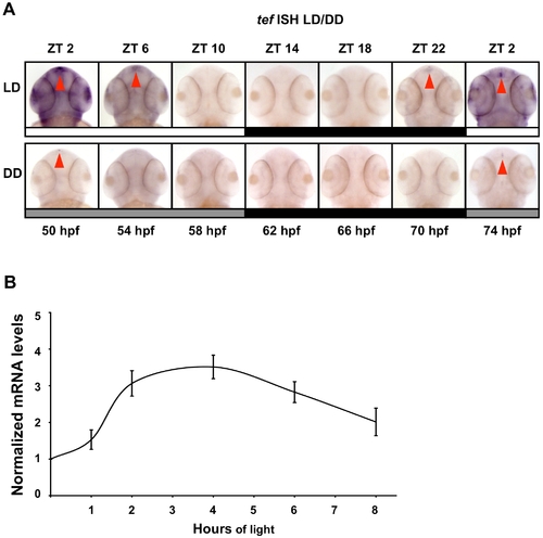
Temporal and spatial expression pattern of TEF under LD and DD cycles. (A) During the first 2 d of development, embryos were exposed to LD cycles. During the third and fourth days of development, embryos were kept under LD or under DD, sampled at 4 h intervals (50–74 hpf), and subjected to whole mount ISH for tef (Genebank Accession number U43671). White bars represent light phase, black bars represent dark phase, and gray bars represent subjective day (ZT, zeitgeber time). Red arrows indicate expression in the pineal gland. Tef is expressed throughout the body and cranial areas with augmented expression in the pineal gland and exhibits a circadian expression pattern with higher levels at the beginning of the subjective day (DD). Under LD, tef expression increases before lights on (ZT 2) and the amplitude of rhythmicity increases. (B) PAC-2 cells were maintained for 5 d in DD. Subsequently, total RNA was extracted from cells kept in darkness or exposed to light for different time periods (1, 2, 4, 6, 8 h). Quantification of tef mRNA levels was performed using qRT-PCR. The mRNA levels in each sample are expressed relative to the level of cells kept in DD. Values shown are the mean from three independent cell pools. Error bars represent SE. These results indicate that tef mRNA levels increase following exposure to light, peaking at 4 h of exposure. Statistical analysis was performed by one sample t-test. All light-treated samples showed significantly higher tef mRNA expression levels relative to DD controls. |
| Gene: | |
|---|---|
| Fish: | |
| Condition: | |
| Anatomical Term: | |
| Stage Range: | Long-pec to Protruding-mouth |

