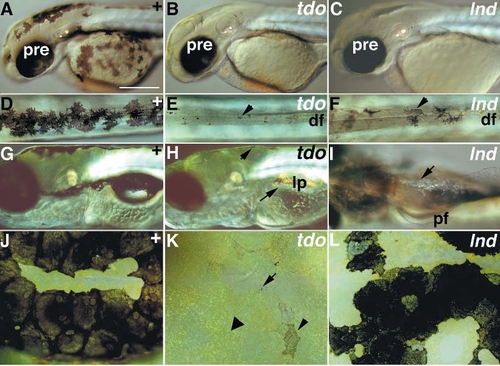
Delayed melanophore differentiation phenotypes (Class VI.B). Wild-type siblings (A,D,G,J) are compared with homozygous mutants for tdo (B,E,H,K) and lnd (C,F,I,L) on the third day (A-F) and the sixth day (G-L). In both tdo and lnd mutants (B and C) the pigmented retinal epithelium is normal, but melanophores are all tiny (tdo, E) or a mixture of tiny and pale cells (lnd, C,F). On the sixth day, tdo mutants now have a few larger, but still pale melanophores (short arrow, H; small arrowhead, K), in addition to tiny melanin spots (arrow, K), although large areas still lack visible melanophores (e.g. none visible around lateral patches: long arrow, H). Xanthophores fill the regions of the dorsal stripe normally occupied by melanophores (large arrowhead, K). In contrast, lnd mutants are now almost fully recovered. Some melanophores are still a little paler (L), and they are still invisible in the yolk sac stripe in some individuals (arrow, I). D-F and J-L are dorsal views of the dorsal stripe in the midtrunk (D-F) or the dorsal head (J-L). I is a ventral view of the trunk. Abbreviations as Fig.2; df, dorsal fin; lp, lateral patch; pf, pectoral fin Scale bars, 250 μm (A-C,G-I) and 100 μm (D-F,J-L).
|
