
Fig. 4
- ID
- ZDB-FIG-091118-14
- Publication
- Schilling et al., 1996 - Jaw and branchial arch mutants in zebrafish. I. Branchial arches
- Other Figures
- All Figure Page
- Back to All Figure Page
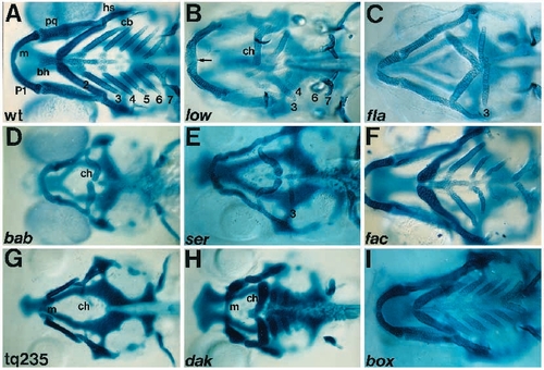
Skeletal defects in mutants (ventral view; pharyngeal arches). (A) Wild type, wt. The seven arch segments contain 13 bilateral and 6 midline cartilages. (B) low. The ceratohyal and ceratobranchials are reduced, P5 is absent. P7, including teeth, is well developed. Meckel’s cartilages fuse in the midline (arrow). (C) fla. All arches are reduced. Meckel’s cartilages are narrow and pointed. Ceratohyals point posteriorly and contact ceratobranchials of P3. There are single teeth on P7. (D) bab. Branchial arches are absent except the basibranchial of P3. Anterior arches are reduced, ceratohyals kinked and posteriorly displaced. (E) ser. Ceratobranchials of P4-P7 are absent; basibranchials are present. Anterior arches are reduced, ceratohyals kinked and posteriorly displaced. (F) fac. Branchial arches, P4-6, are reduced. (G) tq235. Branchial arches are absent. Anterior arches are severely reduced. (H) dak. All arches are abnormally short and wide, narrowing medially. (I) box. All arches are shorter and wider than wt. |