|
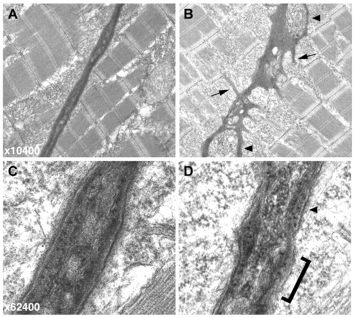
Electron micrographs of the vertical myoseptum reveal a highly irregular MTJ in sof embryos. (A,B) At lower magnification (x10,400), the wild-type myoseptum is compact and linear (A) whereas the sof myoseptum appears severely distorted, even in the absence of fibre detachment (B). Numerous processes extending into the myotome are clearly visible in the mutant (B, arrows), and blisters within the myoseptum are also indicated (arrowheads). (C,D) At higher magnification (x62,400), the smooth, layered structure of the BM at the wild-type MTJ is evident (C). By contrast, the sof BM lacks organisation, with a rough surface (bracketed) and only patches of layering (arrowhead) (D).
|