
Fig. 1
- ID
- ZDB-FIG-090114-16
- Publication
- Ober et al., 1999 - Signals from the yolk cell induce mesoderm, neuroectoderm, the trunk organizer, and the notochord in zebrafish
- Other Figures
- All Figure Page
- Back to All Figure Page
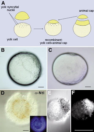
The yolk cell, but not marginal zone tissue, can induce mesoderm in animal cap tissue. (A) Scheme of the experimental procedure. All blastomeres were removed from a high-stage embryo. This yolk cell was then recombined with an animal cap removed from a sibling embryo between high- and sphere-stage. (B) Lateral view of a high-stage embryo, which was incubated in Ca2+-free Ringer’s; animal pole is up. Only the marginal-most blastomeres remain attached to the yolk cell, via tight junctions. (C) Animal view of a completely blastomere-free yolk cell from a high-stage embryo. (D) Animal view of one conjugate. Blastomeres at the periphery of the cap express the pan-mesodermal marker Ntl. (Inset) Animal view of the same conjugate incubated with DAPI showing the external YSL nuclei arranged in a ring-like fashion around the animal cap. The nuclei of the YSL are larger than those of the animal cap blastomeres. (E, F) An animal cap–marginal zone conjugate stained for Ntl protein. The endogenous Ntl expression in the lineage-labeled marginal zone tissue (F) is detected in brown (E), but Ntl expression is not induced in the animal cap explant. Arrowheads point out corresponding nuclei. Scale bars: 100 μm. |
Reprinted from Developmental Biology, 215(2), Ober, E.A. and Schulte-Merker, S., Signals from the yolk cell induce mesoderm, neuroectoderm, the trunk organizer, and the notochord in zebrafish, 167-181, Copyright (1999) with permission from Elsevier. Full text @ Dev. Biol.