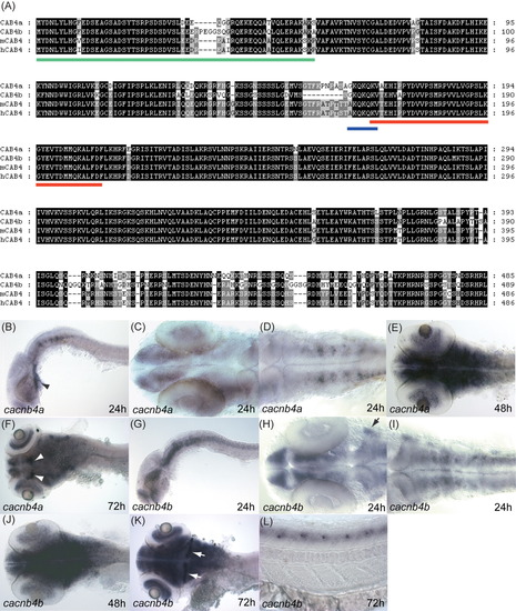
Zebrafish cacnb4a and cacnb4b genes. A: Alignment of protein sequences encoded by zebrafish and mammalian CACNB4 genes shows that they are highly homologous to each other. The red bar underlines the β interaction domain (BID). The blue bar underlines the AKQKQKQ/S/V motif that is conserved in β1, β3 and β4. The green bar underlines the β4-specific D1 domain. B: Lateral view showing cacnb4a expression in the brain and the cardiac tube (arrow) at 24 hpf. C: Dorsal view showing expression of cacnb4a in the forebrain, retina, and midbrain at 24 hpf. D: Dorsal view showing cacnb4a is expressed in the hindbrain and spinal cord at 24 hpf. E: Dorsal view showing cacnb4 is expressed strongly in the brain at 48 hpf. F: Dorsal view of 72 hpf embryos showing cacnb4a expression in the brain. The white arrowheads indicate the two groups of cells in the dorsal midbrain with strong expression of cacnb4a. G: Lateral view showing cacnb4b is expressed in the brain and spinal cord at a higher level than cacnb4a at 24hpf. Note cacnb4b is not detected in the cardiac tube. H: Dorsal view of the hindbrain showing cacnb4b is strongly expressed in the forebrain, midbrain, and trigeminal ganglia (arrow) at 24 hpf. I: Dorsal view showing cacnb4b is expressed in hindbrain and spinal cord at 24 hpf. J: Dorsal view showing strong expression of cancb4b in the brain at 48 hpf. K: Dorsal view of 72 hpf embryo showing expression of cacnb4b persists in the brain. The white arrows indicate stronger expression of cacnb4b in the cerebellum compared with the rest of the brain. L: Lateral view at 72 hpf showing expression of cacnb4b by periodically located cells in the spinal cord. Anterior is left in all the panels.
|

