|
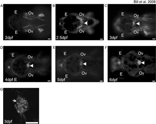
Development of the mCP as defined by EtMn16 larvae.
Fluorescent images of dorsal-oriented live zebrafish taken at 2 dpf (A), 2.5 dpf (B), 3 dpf (C), 4 dpf (D), 5 dpf (E), and 6 dpf (F); arrowhead indicates mCP. GFP-expressing cells first appear diffusely across the roof plate of the fourth ventricle (B), as defined by the level of the otic vesicles (Ov). Cells migrate toward the midline and finish coalescence by 4 dpf. The expression in this structure remains static through 6 dpf. Not all cells of the mCP express GFP (G, arrow). In all images, anterior is to the left, and scale bar is 50 μm. Abbreviations: eye (E), otic vesicle (Ov).
|