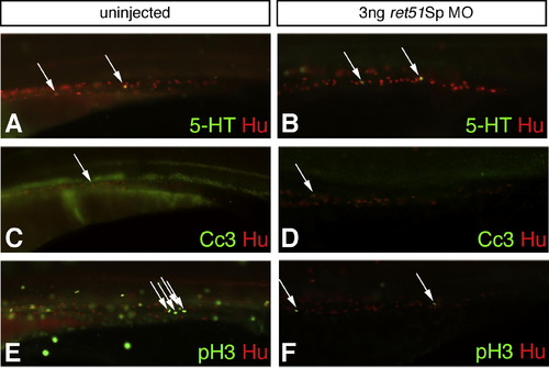
Fish injected with ret51Sp MO analyzed for effects on the serotonergic subpopulation of ENS, on cell death and cell proliferation. Fish injected with ret51Sp MO are compared to uninjected fish by analysis at 4dpf for the presence of Hu-expressing enteric neurons (red, A–F), serotonergic neurons (5-HT positive, green, A–B), cells undergoing cell death (cleaved caspase 3 (Cc3) positive, green, C–D), and proliferative cells (phoshpo-histone 3 (pH 3) positive, green E–F). Equivalent numbers of 5-HT expressing serotonergic neurons are observed in uninjected fish (A, arrows) and those injected with 3 ng ret51Sp MO (B, arrows). Only very small numbers of cells are observed to be undergoing cell death in uninjected (C, arrow) or 3 ng ret51Sp MO injected fish (D, arrow). Significant reductions in numbers of pH 3 positive cells are observed globally in 3 ng ret51Sp MO injected fish (F) as compared to uninjected counterparts (E), and the same trend is observed within the developing gut (E, F, arrows). This difference is cell proliferation likely to reflect the significant non-specific developmental delays observed in ret51Sp MO injected fish, which leads to low levels of survival of fish to stages showing the morphological criteria of a 4dpf fish (Kimmel et al., 1995). It is likely, therefore, that the observed reduction in cell proliferation is largely artefactual and not a reflection of a role for Ret51 in regulating cell proliferation. Images for comparison are photographed at the same magnification.
|
