|
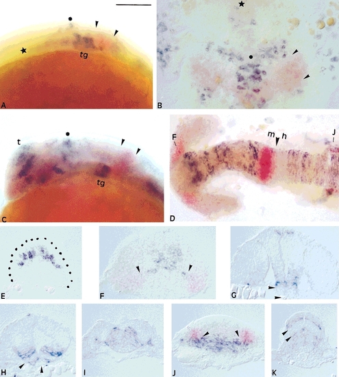
Expression of ngn1 and pax-b during formation of the zebrafish CNS is specific and mutually exclusive. Note the gaps in expression of ngn1 in G,H,K illustrating the layered organization of the neural tube. Small arrowheads define the borders of pax-b expression at the midbrain-hindbrain boundary; the large arrowhead defines the posterior boundary of the midbrain. Asterisks, the dorsal expansion of ngn1 cells; stars, the anterior extent of the floor plate. F,J: Defines positions of cross-sections shown on F and J. A,B,C,D,F,J show two color in situ hybridization: pax-b, magenta, ngn1, blue. E-K: cross-sections; A,C,D: anterior to the left; B: anterior to the top. A,B: 10.5 hpf; C,D: 18 hpf. C,D,F,J: same embryo at 18 hours postfertilization (hpf). E,G,H,I,K: 24 hpf. Scale bar = 100 μm.
|