
FIGURE
Fig. 2
- ID
- ZDB-FIG-080411-5
- Publication
- Wolman et al., 2008 - Transient axonal glycoprotein-1 (TAG-1) and laminin-alpha1 regulate dynamic growth cone behaviors and initial axon direction in vivo
- Other Figures
- All Figure Page
- Back to All Figure Page
Fig. 2
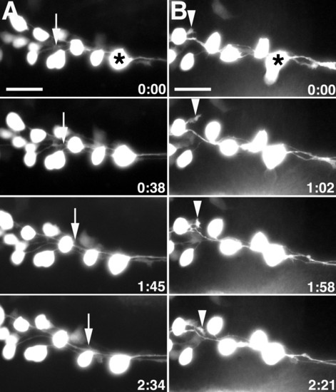
MLF growth cones fasciculate along neighboring MLF axons and are inhibited by surrounding tissue.(a, b) Images from time-lapse sequence of MLF axon outgrowth in uninjected Tg(pitx2c:gfp) embryos. Ventral views, anterior to the left, midline is up. Asterisks denote the caudal-most nucMLF cell. (a) The arrows indicate the position of the growth cone that fasciculates along a neighboring MLF axon. (b) The arrowheads label the growth cone that is repelled by surrounding tissue. The time stamp shows hours: minutes. Scale bar = 25 μm. |
Expression Data
| Gene: | |
|---|---|
| Fish: | |
| Anatomical Term: | |
| Stage: | 14-19 somites |
Expression Detail
Antibody Labeling
Phenotype Data
Phenotype Detail
Acknowledgments
This image is the copyrighted work of the attributed author or publisher, and
ZFIN has permission only to display this image to its users.
Additional permissions should be obtained from the applicable author or publisher of the image.
Full text @ Neural Dev.