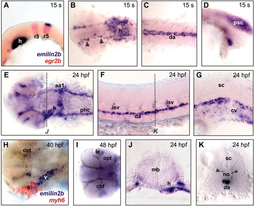
Whole-mount in situ hybridization for emilin2b in zebrafish embryos at different developmental stages. A: Expression in the heart primordium and in the dorsal side of fifth rhombomere of the hindbrain at 15 somites (s), as confirmed by double labeling with a egr2b antisense probe, that marks the third and fifth rhombomeres (in red). B: Expression in blood vessels of the head (arrowheads) and in the heart at 15 s. C: Expression in the dorsal aorta at 15 s. D: Expression in the posterior region of spinal cord at 15 s. E: Labeling of the heart and the circulatory system of the head at 24 hours postfertilization (hpf). F: Lateral view of trunk, showing expression in dorsal aorta and intersegmental blood vessels. G: Expression in the caudal vein at 24 hpf. H: Double hybridization with emilin2b (blue) and myh6 (red) probes, showing expression in the ventricle and part of the atrial chamber of heart at 40 hpf. I: At 48 hpf, emilin2b expression in the circulatory system becomes restricted to head vessels. J: Cross-section of a 24 hpf embryo (level of section is marked by a dotted line in E), showing expression in heart and circulatory system. K: Cross-section of a 24 hpf embryo (level of section is marked by a dotted line in F), showing expression in dorsal aorta and intersegmental blood vessels (arrowheads). All embryos were dissected away from the yolk and mounted between glass coverslips. In A-I, anterior side of embryo is on the left; in cross-sections, dorsal side is at the top. A,D,F and G are lateral views; B and C are dorsal views; E, H. and I are ventral views. aa1, mandibular arch; a, atrium; chf, choroid fissure; cv, caudal vein; da, dorsal aorta; h, heart; isv, intersegmental blood vessels; le, lens; mb, midbrain; no, notochord; opt, optic cup; phc, primordial hindbrain channel; psc, posterior spinal cord; r3, third rhombomere; r5, fifth rhombomere; sc, spinal cord; v, ventricle.
|
