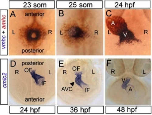
Fig. S1
|
Stages of Zebrafish Heart Development (A-C) Dorsal views of double labeling in situ hybridisation with ventricle myosin heavy chain (vmhc, blue) and atrial myosin heavy chain (amhc, red) probes. After fusion of the bilateral heart fields (23-somites, 20 hours post fertilization hpf) a cardiac disk is formed which is located ventrally to the neural tube and endoderm (A). While future ventricle cells (blue), which are positioned centrally in the disk, start to form a cone the future atrial cells (red), located at the periphery of the disk, remain spread out on the yolk (B). At the same time the venous pole of the heart is displaced towards the left while the arterial pole of the tube remains positioned at the midline, a process referred to as cardiac jogging. Slightly later, when extension of the tube has proceeded, also the atrial cells are incorporated into the linear heart tube, which is clearly jogged towards the left. D) Dorsal view of a similar stage embryo as shown in (C). Anterior and posterior have been reversed to allow better comparison with succeeding looping stages during which the heart becomes located more anteriorly due to extension of the embryo. E,F) Anterior views of looping stage embryos. At the onset of rightward looping a Cloop is formed during which the atrioventricular canal becomes positioned at the midline (E). When looping has completed a S-shaped heart is formed with the ventricle positioned to the right and the atrium to the left (F). AVC, atrioventricular canal, A atrium, hpf hours post fertilization, IF inflow, L left, OF outflow, R right. |
| Genes: | |
|---|---|
| Fish: | |
| Anatomical Terms: | |
| Stage Range: | 20-25 somites to Long-pec |
Reprinted from Developmental Cell, 14(2), Smith, K.A., Chocron, S., von der Hardt, S., de Pater, E., Soufan, A., Bussmann, J., Schulte-Merker, S., Hammerschmidt, M., and Bakkers, J., Rotation and asymmetric development of the zebrafish heart requires directed migration of cardiac progenitor cells, 287-297, Copyright (2008) with permission from Elsevier. Full text @ Dev. Cell

