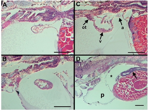
Fig. S2
|
Histological analysis of 4 dpf wt1MO embryos. The hearts of embryos injected with the wt1 Morpholino are morphologically normal. (A) Atrium (arrow) from a wt1MO embryo. The chamber is single-layered and continuous with the sinus venosus. (B) Ventricle from a wt1MO embryo. Note the thicker ventricular wall as compared to the single-layered atrium in (A). (C) Sagittal section through a wt1 morpholino-injected embryo showing both the single-layered atrium (a) and thicker ventricular layer (v) and the outflow tract (ot) in a single section. (D) Lower magnification view of a wt1MO embryo with the pericardial edema marked by (p) and coelomic edema marked by and asterisk. The gut appears to be well differentiated (arrow). All sections shown are in the sagittal plane with anterior to the left, scale bar is 100 microns in all panels. |
Reprinted from Developmental Biology, 315(1), Serluca, F.C., Development of the proepicardial organ in the zebrafish, 18-27, Copyright (2008) with permission from Elsevier. Full text @ Dev. Biol.

