
Fig. 2
- ID
- ZDB-FIG-080114-12
- Publication
- Rieger et al., 2008 - Polysialyltransferase expression is linked to neuronal migration in the developing and adult zebrafish
- Other Figures
- All Figure Page
- Back to All Figure Page
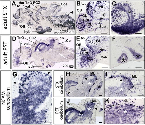
Comparison of stx and pst expression in the adult zebrafish brain. All panels show medial sagittal sections, anterior is to the left. A-C,H,I show stx expression and D-F,J,K show pst expression. A: Overview of stx expression. D: Overview of pst expression. B,E: Cells in the pallial migratory domains of the telencephalon and olfactory bulb are marked by asterisks. C,F: Ventricular staining in the hypothalamic region. G: ncam expression in the molecular layer (ML), Purkinje cell layer (PCL, black arrowhead), and granule cell layer (GCL, white asterisk) of the cerebellum. H: Higher magnification of the cerebellum showing stx expression in some cells of the ML and strong expression in cells of the PCL, extending from the corpus into the valvula cerebelli (black arrowheads). Weak expression levels are present in the caudal lobe (black asterisk) and in the GCL (white asterisk). I: A higher magnification of stx expression in the adult cerebellum. J: Strong pst expression in the GCL of the corpus cerebelli and in the valvula cerebelli being more pronounced than stx expression (H, white asterisk). K: A higher magnification of pst expression in the adult cerebellum, being detected in the ML and GCL, separated by a dashed line. Cc, crista cerebellaris; Cce, corpus cerebelli; ECL, external cellular layer; GL, glomerular layer; hyth, hypothalamus; IL, internal cellular layer; med, medulla; ML, molecular layer; OB, olfactory bulb; Pa, pallium; PGZ, periventricular gray zone of the optic tectum (TeO); pn, preoptic nucleus; Sub, subpallium; tc, telencephalon; tha, thalamus; val, valvula cerebelli; vl, vagal lobe. Scale bars = 200 μm. |