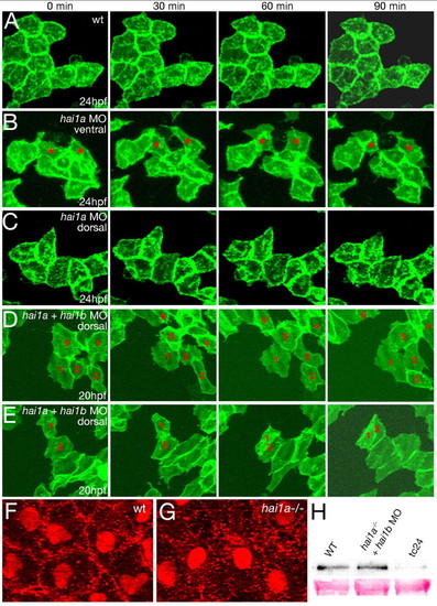
Loss of Hai1 activity abrogates the epithelial properties of basal keratinocytes. (A-E) Stills of in vivo time-lapse recordings of clusters of basal keratinocytes labeled with membrane-bound GFP, at the indicated times after 24 hpf (A-C) or 20 hpf (D,E); wild-type sibling (wt; A; see Movie 1 in the supplementary material); hai1a morphants (B,C; see Movies 2 and 3 in the supplementary material); and hai1a + hai1b double morphants (D,E; see Movies 4 and 5 in the supplementary material). Epidermal cells in the ventral regions of a hai1a morphant (B) display a mesenchymal-like behavior (examples indicated by asterisks; recorded region indicated in Fig. 6G). By contrast, epidermal cells in more-dorsal/posterior regions (C) form a rigid epithelium, as in wild-type embryos (A). In embryos lacking both Hai1a and Hai1b, motility and fibroblastoid behavior of basal keratinocytes is further enhanced, evident much earlier and now also seen in dorsal trunk/tail regions (D; individual keratinocytes labeled with numbers). Often, cells migrate on top of each other (E; cells 1 and 2). (F,G) Ventral confocal views of yolk epidermis of a 24 hpf wild-type sibling (F) and a hai1a mutant embryo (G), with immunofluorescent detection of E-cadherin (E-cad) and p63, marking nuclei of basal keratinocytes. (H) Anti-E-cad western blots of embryonic extracts from 24 hpf wild-type siblings (left lane) and hai1a mutants injected with hai1b MO (middle lane), revealing unaltered E-cad protein levels in double-deficient embryos. By contrast, offspring of smad5dtc24 heterozygous mothers (Hild et al., 1999), which, according to anti-p63 immunostainings, lack basal keratinocytes (data not shown), display strongly reduced E-cad protein levels (right lane).
|
