
Fig. 4
- ID
- ZDB-FIG-070102-4
- Publication
- Gregg et al., 2003 - Positional cloning of the young mutation identifies an essential role for the Brahma chromatin remodeling complex in mediating retinal cell differentiation
- Other Figures
- All Figure Page
- Back to All Figure Page
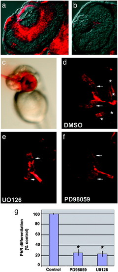
Disruption of MAP kinase activity blocks retinal differentiation. (a and b) Transverse retinal sections showing dual phosphorylated ERK 1/2inWT (a) and the lack of retinal activation of MAP kinase in yng mutant (b) eyes. (c) Example of focal intraocular injection. MAP kinase kinase (MEK) 1/2 inhibitors were injected intraocularly at 42 hpf, a time when ganglion cells have differentiated, but before photoreceptor differentiation. Ganglion cell and photoreceptor markers were assessed at 60 hpf. Phenol red is used as a visible marker to monitor injections. (d–f) Retinal sections of embryos injected with DMSO control (d) or MEK 1/2 inhibitors U0126 (e) and PD98059 (f) and then processed for a mixture of markers that indicate ganglion cell differentiation (zn8, arrows) and photoreceptor differentiation (zpr1, *). Note the absence of photoreceptor differentiation in embryos treated with either U0126 or PD98059. Ganglion cell markers were not affected, demonstrating the inhibitors did not alter retinal cell survival. (g) Quantitative comparison of photoreceptor differentiation between embryos with intraocular injections of DMSO, U0126, or PD98059. After statistical comparison, data were transformed to percent of control. *, P < 0.001, two-tailed Student′s t test; n = 8. |