|
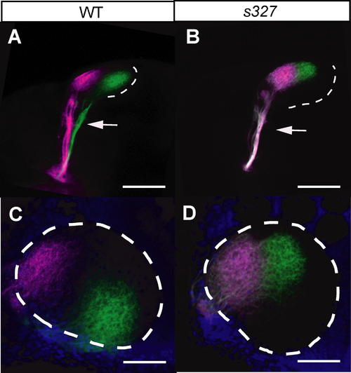
The darl Mutant Shows Retinotectal Mapping Deficits. (A and B) The nasal-dorsal quadrant of the retina was labeled with DiO (green), and the temporal-ventral quadrant was labeled with DiD (magenta). In darls327, the ventral branch of the optic tract is missing (arrow). Scale bar is 100 μm. (C and D) Dorsal view of the tectum in the same larvae as in A and B. The ventral half of the darls327 tectum is not innervated by the dorsal-nasal RGC axons. Anterior is to the left and ventral is to the bottom. Tectal neuropil is demarcated by the dotted line, based on DAPI counterstaining (blue). Scale bar is 50 μm.
|

