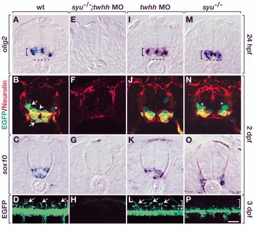
Motoneurons and oligodendrocytes have genetically separable requirements for Hh signaling. Top three rows of panels show transverse sections through trunk spinal cord, with dorsal upwards. Bottom row shows lateral views of intact olig2:egfp embryos, with dorsal upwards and anterior towards the left. (A) Wild-type embryos expressed olig2 RNA in the ventral spinal cord (bracket), but the ventralmost cells were olig2–. Broken line indicates the ventral boundary of the spinal cord. (B) olig2:EGFP+ cells of wild-type embryos included Neurolin+ SMNs, OPCs (arrows), Neurolin– cells near ventricle (arrowhead) and a faint GFP+, Neurolin– cell near pial surface (asterisk), which could be a primary motoneuron or VeLD interneuron. (C) sox10+ OPCs in wild-type embryo. (D) Arrows indicate dorsally migrated OPCs in wild-type embryo. Bracket indicates dorsal spinal cord. (E-H) syu–/– embryos injected with twhh MO lacked olig2+ cells (E), secondary motoneurons (F) and OPCs (G,H). (I-L) Wild-type embryos injected with twhh MO expressed olig2 RNA (I) and produced secondary motoneurons (J) and OPCs (K,L). syu–/– embryos expressed olig2 RNA (M), but olig2+ cells were located more ventrally than normal. (M-P) syu–/– embryos had olig2:EGFP+, Neurolin+ secondary motoneurons and olig2:EGFP+, Neurolin– cells (asterisks) (N); however, they did not have OPCs (O,P). (O) sox10+ cells are probably Schwann cells (Dutton et al., 2001), which do not appear in all sections. Scale bar: 20 µm for upper three rows; 40 µm for bottom row.
|
