- Title
-
Midline signalling is required for Pax gene regulation and patterning of the eyes
- Authors
- Macdonald, R., Barth, K.A., Xu, Q.L., Holder, N., Mikkola, I., and Wilson, S.W.
- Source
- Full text @ Development
|
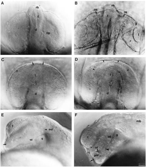
Development of the eye in living wild-type embryos and embryos homozygous for the cyclops mutation. (A,B) Wildtype embryos, (C-F) cyclops embryos. (A) 13s, dorsal view. (B) 24 hour, dorsal view. The arrow indicates the rostral limit of optic cup invagination. The position of the telencephalon is indicated although it is above the plane of focus. (C) 13s, dorsal view. The arrows indicate the bridge of fused retina around the anterior pole of the neural keel. The anterior hypothalamus (see A and B) is missing from mutant embryos. (D) 20-22s, dorsal view. The arrowheads indicate minor invagination in the bridge of fused neural retina. (E) 16s, lateral view focused on the midline. The arrow indicates the area of prospective retinal fusion. A large gap is present beneath the diencephalon. (F) 20-22s, lateral view focused on the midline. The arrow indicates a small amount of invagination in the bridge of prospective retinal tissue. The arrowhead indicates the caudal limit of retinal fusion. Abbreviations: d, diencephalon; g, gap in mid-diencephalic neuroepithelium; l, lens; mb, midbrain; md, mid-diencephalon; ol, optic lumina; op, optic primordia; os, optic stalks; pnr, presumptive neural retina; r, retina; rh, rostral hypothalamus; t, telencephalon; vd, ventral diencephalon. Scale bar, 25 μm. PHENOTYPE:
|
|
Pax protein distribution in wild-type and cyclops mutant embryos. Whole-mounted embryos labelled with anti-Pax6 antibody (A-F), or labelled with anti-Pax2 antibody (G,H). (A,B) Dorsal views of 15-16s wild-type and cyclops embryos respectively. The arrowheads in B indicate the bridge of rostral retinal fusion. (C,D) Dorsal and lateral (focused on the midline) views respectively of 20s cyclops embryos. The arrowheads indicate the area of retinal fusion in C. In D the lateral portions of the cyclopic eye have been removed. The arrowhead indicates the caudal limit of eye fusion. (E) Frontal view of 28s cyclops mutant embryo. (F) Lateral view of 28 hour cyclops mutant embryo also labelled with anti-tubulin antibody (black labelling of axons). This embryo was also examined in a different study (Macdonald et al., 1994). (G,H) 18- 20s dorsal views of wild-type (G) and cyclops (H) embryos labelled with anti-Pax2 antibody. The arrow in H indicates a few cells near the midline weakly labelled with anti-Pax2 antibody. Abbreviations: d, diencephalon; hb, hindbrain; mb, midbrain; mdf, mid-diencephalic furrow; l, lens; mt, midbrain tegmentum; ne, neural ectoderm; nr, nasal retina; os, optic stalks; np, nasal placode; op, optic primordia; ov, optic veiscle; ple, presumptive lens ectoderm; pnr, presumptive neural retina; ppe, presumptive pigment epithelium; se, surface ectoderm; t, telencephalon; te, tectum; tr, temporal retina; vd, ventral diencephalon. Scale bars, 50 μm. EXPRESSION / LABELING:
|
|
rtk2 and msxC expression in wild-type, cyclops and shh-injected embryos. Whole-mounted embryos with rostral to the left (A,B,F-H) or up (D,E). (A,B) Lateral views of rtk2 expression in 24 hour wild-type (A) and cyclops mutant (B) embryos. The arrowheads indicate the boundary between nasal and temporal retina at the posterior groove. The asterisk and filled circle indicate equivalent positions of the eye in B and C. (C) Frontal view of rtk2 expression in a 20s cyclops mutant embryo. rtk2 expression in temporal retina is continuous around the rostral pole of the brain. (D,E) Dorsal views of rtk2 expression in 24 hour wild-type and shh-injected embryos respectively. rtk2 expression is widespread thoughout the retinal tissue in the shh-injected embryo. (F) Lateral views of msxC expression in the eye (arrowheads) of wild-type (right) and cyclops mutant (left) 18s embryos. (G,H) Lateral views of msxC expression in the eyes of shh injected (right) and wild-type (left) 18-20s (G) and 24 hour (H) embryos. In addition to an absence of msxC expression in the eyes (arrowheads), msxC is reduced throughout the head (arrows). Abbreviations: cf, choroid fissure; d, diencephalon; l, lens; nr, nasal retina; os, optic stalk; t, telencephalon; te, tectum; tr, temporal retina. Scale bars, 50 μm. EXPRESSION / LABELING:
|
|
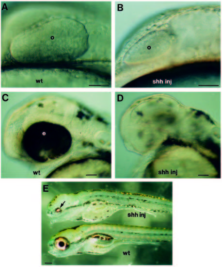
Development of the eye in living wild-type embryos and embryos injected with shh RNA. Lateral views with rostral to the left. (A,B) 10-12s, wild-type and injected embryos respectively. (C,D) 48 hour, wild-type and injected embryos respectively. The injected embryo has virtually no pigment epithelium and very little neural retina. (E) Comparison of injected (upper) and wild-type (lower) embryos at 3-4 days of development. The arrow indicates the much reduced retina in the injected embryo. Abbreviations: e, eye; o, optic primodium. Scale bars, (A-D) 50 μm; (E) 200 μm. |
|
shh has opposite effects upon Pax2 and Pax6 expression in the optic primordia. Whole-mounted 16-18s embryos labelled with antibodies to Pax6 (A-C) or Pax2 (D-F), or hybridised with antisense RNA probes to pax6 (G,H) or pax2 (I,J). Rostral CNS is to the left. (A) Dorsal views of Pax6 distribution in shh-injected (upper) and wild-type (lower) embryos. (B,C) Higher magnification views showing Pax6 distribution in the forebrain and eyes of wildtype (B) and injected (C) embryos. Some anti-Pax6 antibody labelling persists in the caudal portions of the optic primordia of the injected embryo (arrowheads in C). (D) Dorsal view of Pax2 distribution in intact wild-type (upper) and injected (lower) embryos. (E,F) Higher magnification views showing Pax2 distribution in the forebrain and optic primordia of wild-type (E) and injected (F) embryos. The most caudal cells of the optic primordia of the injected embryo are not labelled with the anti-Pax2 antibody (arrowheads in F). The area of attachment of the optic primordia to the diencephalon is greatly increased in the shh-injected embryo (double headed arrow in F). (G,H) Dorsal view of pax6 expression in the eye primordia of wild-type (G) and shh-injected (H) embryos. (I,J) pax2 expression in the optic primordia of wild-type (I) and shh-injected (J) embryos. Abbreviations: d, diencephalon; l, lens; nr, neural retina; os, optic stalk; pe, pigment epithelium. Scale bars, 50 μm. |
|
Altered Pax protein distribution correlates with abnormal segregation of the optic primordia into optic stalks and retinae in embryos injected with shh RNA. Rostral is to the left in A-C and F. All embryos in this figure showed only moderate reduction in the extent of the retinal tissue. Embryos are labelled with anti-Pax2 (A-F), HNK1 (G) or anti-Pax6 (H) antibodies. (A) Lateral views of 24 hour injected (left) and wild-type (right) embryos. The arrowheads indicate sites of Pax2 expression at the midbrain/hindbrain boundary, in the otocysts, in hindbrain neurons and in the nephric duct. (B,C) Ventral views of wild-type (B) and injected (C) embryos. (D,E) Frontal views of wild-type (D) and injected (E) embryos. The arrows in E indicate the expansion of Pax2 expression in the optic stalk region of the injected embryo. (F) Horizontal section through the retina of an shh-injected embryo at 2 days of development. The distal extent of Pax2 distribution (arrowheads) is coincident with the proximal limit of pigment epithelium formation. Most of the cells in the deeper layers of the retina still contained Pax6 at this stage as judged by anti-Pax6 labelling of adjacent serial sections. (G,H) Frontal sections through the retina of an shhinjected 5-day old fish labelled with HNK1 antibody (G; brown labelling of axons) or anti-Pax6 antibody (H). At this stage Pax6 protein is restricted mainly to amacrine cells (Macdonald and Wilson, unpublished observations). The antibody labelling reveals relatively normal lamination of the neural retina. The arrows indicate medial regions of the neural retina over which there is no pigment epithelium. Abbreviations: a, amacrine cell layer; g, ganglion cell layer; hy, hypothalamus; inl, inner nuclear layer; ipl, inner plexiform layer; l, lens; nr, neural retina; os, optic stalk; p, photoreceptor layer; pe, pigment epithelium; t, telencephalon. Scale bars, 50 μm. |


