
- Title
-
Esketamine Exposure Impairs Cardiac Development and Function in Zebrafish Larvae
- Authors
- Huang, S., Wang, J., Lin, T., He, C., Chen, Z.
- Source
- Full text @ Toxics
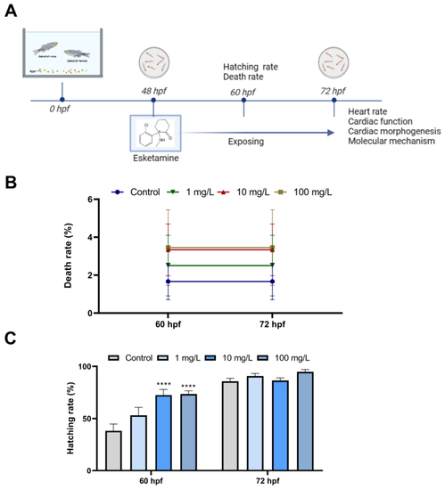
Developmental effects of zebrafish embryos exposure to esketamine. (A) Scheme of experiment design. (B) Mortality rate from 60 to 72 hpf. (C) Hatching rate from 60 to 72 hpf A two-way analysis of variance (ANOVA) was employed for the statistical analysis of the death and hatching rates. This was followed by multiple comparison tests, and the results were expressed as the mean ± SEM (n = 4). **** p < 0.0001 |

Cardiac function of zebrafish larvae exposed to esketamine at 72 hpf. (A) Morphological alterations were observed using microscopy (Scale bar: 100 µm), (B) heart rate, (C) end-diastolic volume (EDV), (D) end-systolic volume (ESV), (E) stroke volume (SV), and (F) cardiac output (CO). The data underwent a one-way analysis of variance (ANOVA) and were further evaluated using Tukey’s multiple comparisons test. Results are reported as the mean ± SEM (n ≥ 25). * p < 0.05, ** p < 0.01, *** p < 0.001 |
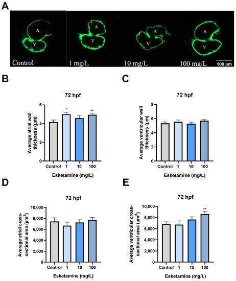
Cardiac morphogenesis of zebrafish larvae exposed to esketamine at 72 hpf. (A) Heart structure visuals showcase the cardiac morphogenesis in larvae exposed to esketamine. The measurements include (B) the average thickness of the atrial wall, (C) the average thickness of the ventricular wall, (D) the average cross-sectional area of the atrium (noted as A for atrium and V for ventricle), and (E) the mean cross-sectional area of the ventricles. Following the statistical analysis with a one-way analysis of variance (ANOVA) and a Tukey multiple comparisons test, the results are reported as the mean ± SEM (n = 10). * p < 0.05, ** p < 0.01. |

The expression levels of cardiac development-related genes in zebrafish larvae exposed to esketamine at 72 hpf. (A) nkx2.5, (B) gata4, (C) tbx5, and (D) myh6. The data underwent a one-way analysis of variance (ANOVA) and a Tukey multiple comparisons test. The findings are expressed as the mean ± SEM (n = 3). * p < 0.05, ** p < 0.01, *** p < 0.001. |

The expression levels of the calcium signaling pathway genes in zebrafish larvae exposed to esketamine at 72 hpf. (A) ryr2a, (B) ryr2b, (C) atp2a2a, (D) atp2a2b, (E) slc8a3, (F) slc8a4a, and (G) cacna1aa. The expression values of these genes were normalized using gapdh. The evaluation of the data was carried out using Tukey’s multiple comparisons test, and the findings are represented as the mean ± SEM (n = 3). * p < 0.05, ** p < 0.01, *** p < 0.001 and **** p < 0.0001. |

The expression levels of NMDAR-related genes in zebrafish larvae exposed to esketamine at 72 hpf. (A) grin1a, (B) grin1b, (C) grin2ca, and (D) grin2bb. The data underwent a one-way analysis of variance (ANOVA) and a Tukey multiple comparisons test. The findings are expressed as the mean ± SEM (n = 3). |

The level of Ach and activities of AchE and ChAT in zebrafish larvae exposed to esketamine at 72 hpf. (A) Ach. (B) AchE. (C) ChAT. The analysis of the data was performed using Tukey’s multiple comparisons test, and the results were expressed as the mean ± SEM (n = 3). * p < 0.05, ** p < 0.01, *** p < 0.001 and **** p < 0.0001. |
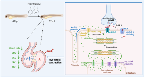
The proposed mechanism of esketamine affects the contraction of cardiomyocytes in zebrafish. Initially, esketamine exposure increased the concentration of Ach, which inhibits calcium channels, resulting in a decrease in the mRNA expression of cacna1aa, which are L-type voltage-gated calcium channels, leading to a decrease in the influx of extracellular calcium ions into the cytoplasm. As a result, the activation of the calcium channel RyR2 is delayed, requiring the attachment of calcium ions to the membrane of the sarcoplasmic reticulum (SR). Additionally, the mRNA expression of ryr2a and ryr2b also decreased after exposure. These combined effects severely impede the release of calcium from the SR in cardiac myocytes, resulting in a relative decrease in cytoplasmic calcium concentration. This relative decrease in calcium concentration disrupts the binding of calcium to myosin, limiting the sliding of myofilaments and ultimately impairing the contractile ability of cardiac myocytes. |