FIGURE SUMMARY
Title

Sleep pressure modulates single-neuron synapse number in zebrafish

Authors
Suppermpool, A., Lyons, D.G., Broom, E., Rihel, J.
Source
Full text @ Nature

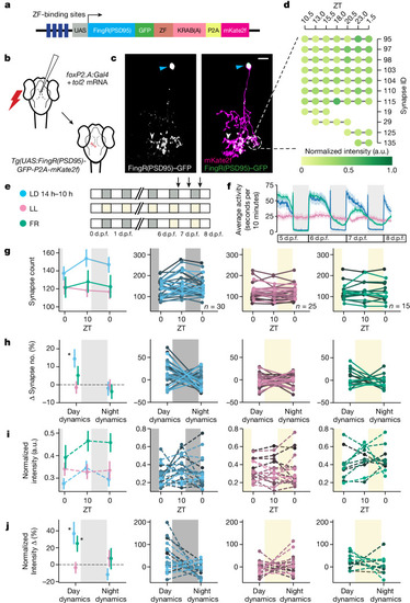
Single-neuron synapse tracking across day–night cycles reveals diverse dynamics.

a, The synapse labelling construct. Zinc finger (ZF) and KRAB(A) domains limit overexpression25. b, The strategy to sparsely label synapses of FoxP2.A+ tectal neurons (Methods). c, Example FoxP2.A:FingR(PSD95)+ neuron at 7 d.p.f., with the synapses (white arrowheads, left), nucleus (blue arrowheads, left) and membrane (magenta, right) co-labelled. d, Overnight time-lapse tracking of select synapses from the neuron in c. The normalized GFP intensity (shading) is shown for each synapse (rows). The complete neuron map is shown in Extended Data Fig. 2a. e, Larvae were raised on 14 h–10 h light–dark (LD) cycles (blue), constant light (LL, pink) or switched from LD to LL at 6 d.p.f. (free running (FR), green), and then imaged (arrows) (Methods). f, The average locomotor activity and 95% confidence intervals (CIs) of larvae reared under LD (blue, n = 75), clock-break LL (pink, n = 84) or FR (green, n = 98) conditions. gj, The mean and 68% CI (column 1) and individual neuron (columns 2–4) synapse counts (g), percentage change in synapse number calculated within each neuron (h), normalized synapse intensity (i) and percentage change in synapse intensity (j) under the LD (blue), LL (pink) or FR (green) conditions. For columns 2–4, a line is shown for each neuron, collected across 8 LD, 4 LL and 4 FR independent experiments. For h, synapse number change (Δ synapse number) dynamics are different during the day from those during the night under LD conditions (*P = 0.043, repeated-measures analysis of variance (ANOVA)). Synapse number change dynamics under LD cycling are significantly different from those under LL conditions (*P = 0.015, main effect of condition, two-tailed mixed ANOVA, post hoc Benjamini–Hochberg correction; Hedge’s g = 0.761). For j, day–night dynamics are significantly different under LD from those under the other conditions (P < 0.01, repeated-measures ANOVA). Both daytime FR and LD day–night dynamics are significantly different from those under the LL condition (mixed ANOVA interaction (condition × time), P = 0.029; FR versus LL, P = 0.038, g = 0.937; LD versus LL, P = 0.027, g = 0.792; post hoc Benjamini–Hochberg correction, two-tailed). At night, LD versus FR, g = −0.538; LD versus LL, g = −0.527. The diagram in a is adapted from ref. 27, CC BY 4.0, and the diagram in b is adapted from ref. 33, CC BY 4.0. The colour key in e applies also to fi.

Source Data

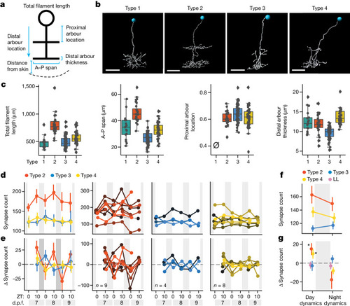
Subtype-specific synapse changes in FoxP2.A tectal neurons over 3 days.

a, The morphological parameters used to characterize FoxP2.A tectal neurons. A–P, anterior–posterior. b, Examples of each morphological subtype, chosen from n = 17 (type 1), n = 28 (type 2), n = 61 (type 3) and n = 42 (type 4) neurons collected over 26 independent experiments. The blue circles label nuclei. c, Example of the parameters used to distinguish the four subtypes. For the box plots, the centre lines show the median, the box limits show the interquartile range and the whiskers represent the distribution for each parameter. The slashed zero indicates that the feature is absent. See also Extended Data Fig. 5. dg, Synapse counts across multiple LD cycles for FoxP2.A tectal neurons of different subtypes. d,e, Average (68% CI) synapse counts (d) and average (68% CI) synapse number change (e) of subtypes (column 1) and for each neuron (columns 2–4), collected over 8 independent experiments. f,g, Average (68% CI) synapse counts (f) and net change (g), averaged across all days and nights for each subtype and larvae, including additional neurons tracked over a single day (Extended Data Fig. 5). Tectal subtype influences synapse changes (mixed ANOVA, interaction P = 0.012, subtype × time). Type 2 (n = 16) and type 4 (n = 15) neurons gain more synapses during the day under LD conditions compared with under LL clock-break conditions (P = 0.018, g = 0.952; P = 0.021, g = 0.812, respectively). At night, both type 2 and type 4 neurons lose synapses relative to type 3 (type 2 versus type 3, P = 0.038; g = −0.714; type 4 versus type 3, P = 0.038, g = −0.781, post hoc Benjamini–Hochberg correction, one-tailed). For b, scale bars, 10 μm.

Source Data

Synapse counts of neurons are modulated by sleep and SD.

a, The 4 h gentle handling SD paradigm (ZT14–ZT18). Larvae were video-tracked and neurons were periodically imaged (arrows). b, The mean ± s.e.m. change in synapse counts per hour for the SD (orange, n = 31 neurons) and control (blue, n = 28) groups. c, Sleep time versus the change in synapse counts per hour for each larva during either the early (ZT14–ZT18, left) or late (ZT18–ZT24, middle) night for controls and after SD (ZT18–ZT24, right). The rate of synapse change is negatively correlated with sleep time during both early and late night but not after SD. d, In control larvae, the change in early night synapse counts is negatively correlated with late night synapse change. Early and late sleepers are defined as larvae that either sleep more in the first or second phase of the night, respectively. e, Synapse counts per hour for early- and late-night sleeping control larvae in the early (ZT14–ZT18) and late (ZT18–ZT24) phases of the night. Data are mean ± s.e.m. fh, The reticulospinal neuron synapse number is modulated by sleep and wake states. f, Example reticulospinal neurons from the Tg(pvalb6:KALTA4)u508 line co-labelled by FingR(PSD95)–GFP (green, nuclei and synapses) and mKate2f (magenta, membrane). Vestibulospinal (VS) and MiD2cm neurons are indicated by the dashed ovals. g, Vestibulospinal (top) and MiD2cm (bottom) neurons from different larvae showing FingR(PSD95)+ synapses (green) co-localized to the cell membrane (magenta). h, Changes in synapse number (mean and 68% CI) from ZT14 to ZT18 for vestibulospinal and MiD2cm neurons. Each dot represents the average across multiple neurons per larva. For b and e, statistical analysis was performed using two-tailed mixed ANOVA interaction (condition × time) with post hoc Benjamini–Hochberg correction; ****P = 0.00007, ***P = 0.0002 and **P = 0.006 (b) and *P = 0.01 (e). For h, statistical analysis was performed using one-tailed Student’s t-tests; *P < 0.03. Scale bars, 15 μm (f) and 10 μm (g). The lines in c and d depict the linear regression with the 95% CI.

Source Data

Single-neuron synapse loss during sleep is driven by boosting adenosine and blocking noradrenaline.

a, Larvae were temporarily treated with sleep-promoting drugs during the day (ZT5–ZT10). The black arrows indicate the imaging periods before and after drug treatment. b, Drug-induced sleep during the day disentangles sleep pressure (that is, low) from sleep amount (that is, high), which are otherwise tightly correlated. c, Drug-treated larvae sleep significantly more during the day compared with the dimethyl sulfoxide (DMSO)-treated controls. d, During the day (from ZT5–ZT10), synapse counts increase under all control and drug conditions, except during co-administration of clonidine and 2-chloroadenosine, when synapses are significantly lost. Data are mean ± s.e.m. n values represent the number of neurons (top row) or fish (bottom row). For c and d, statistical analysis was performed using Kruskal–Wallis tests with post hoc Dunn’s multiple-comparison test (left) and one-way ANOVA (right); not significant (NS), P > 0.5; *P = 0.034, **P < 0.01, ****P < 0.0001.

Source Data

Acknowledgments
This image is the copyrighted work of the attributed author or publisher, and ZFIN has permission only to display this image to its users. Additional permissions should be obtained from the applicable author or publisher of the image. Full text @ Nature