
- Title
-
Establishment of a graphene quantum dot (GQD) based steroid binding assay for the nuclear progesterone receptor (pgr)
- Authors
- Hossain, M.F., Hossain, S., Sarwar Jyoti, M.M., Omori, Y., Ahamed, S., Tokumoto, T.
- Source
- Full text @ Biochem Biophys Rep
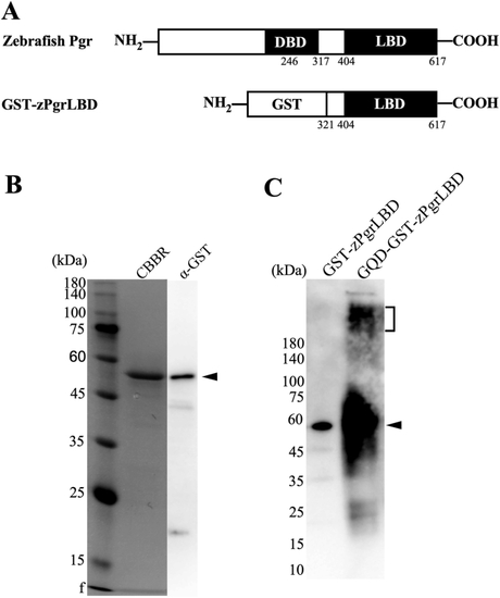
Expression and purification of the ligand-binding domain (LBD) of zebrafish Pgr as a fusion protein with GST and production of GQD-coupled GST-zPgrLBD. A. A diagram of the produced recombinant GST-fusion zPgrLBD. The predicted ligand-binding domain at the |

Fluorescence characteristics of GQD-GST-zPgrLBD and competition of binding of P4-BSA-FITC with GQD-GST-zPgrLBD by steroids and their analogues. (A) The fluorescent scanning pattern of free GQD-GST-zPgrLBD is indicated in blue line. Fluorescent scanning pattern of the reaction mixture with (orange line) or without free P4 (green line). The difference of fluorescence at 520 nm caused by the addition of free P4 is indicated by the double-sided arrow. (B) The dose-dependent effects of steroids (progesterone (P4), 17β-estradiol (E2), testosterone (T), cortisol) and their analogues (17,20β-DHP and Org OD 02) were determined using the established assay. An assay was performed in triplicate for each compound. (For interpretation of the references to colour in this figure legend, the reader is referred to the Web version of this article.) |
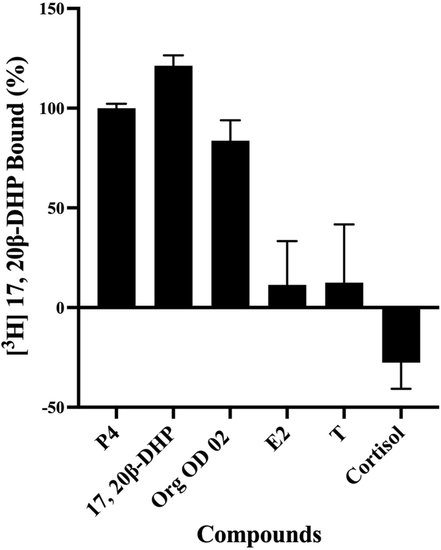
Competition by steroids and progestins for binding to GQD-GST-zPgrLBD. Samples were incubated with 2 nM [3H]-17,20β-DHP and 1 μM of competitor. An assay was performed in triplicate for each competitor. The average of the three assays is indicated with standard deviation. Competition for [3H]-17,20β-DHP binding are expressed as a percentage of maximum specific progesterone (P4) binding. The competitors analyzed were progesterone (P4); 17,20β-DHP; Org OD 02; 17β-estradiol (E2); testosterone (T); Cortisol. |