FIGURE SUMMARY
Title

Spermidine treatment: induction of autophagy but also apoptosis?

Authors
Watchon, M., Wright, A.L., Ahel, H.I., Robinson, K.J., Plenderleith, S.K., Kuriakose, A., Yuan, K.C., Laird, A.S.
Source
Full text @ Mol. Brain

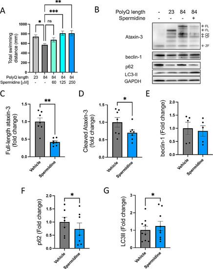
Spermidine treatment alleviates motor dysfunction in transgenic MJD zebrafish together with signs of autophagy induction. (A) Mutant ataxin-3 zebrafish (84Q) were treated with spermidine (60–250 µM) for 5 days and motor behaviour was measured at 6 dpf. Vehicle treated mutant ataxin-3 larvae swam shorter distances compared to the non-transgenic control and spermidine treatment rescued this phenotype (***p < 0.001 and *p < 0.05, n = 96–140). (B) Spermidine treated mutant ataxin-3 larvae were subjected to immunoblotting (B). Densitometric analysis of (C) full-length and (D) cleaved- human ataxin-3 revealed a significant decrease in ataxin-3 expression in spermidine treated EGFP-ATXN3 84Q fish compared to the vehicle treated mutants (** p = 0.002 and p = 0.020 respectively, n = 8–10). Various substrates of the autophagy pathway showed indications of positive autophagic flux. (E) No difference was found in levels of beclin-1, (F) p62 was found to be significantly decreased (*p = 0.0432) and (G) LC3II was found to be increased. (p = 0.0426, all n = 8–10). A one-way ANOVA followed by Tukey post hoc analysis and a paired student t-test was utilised for statistical analysis. Data represents mean ± SEM. Data points reflected in C-G are experimental treatments of approximately 20–25 larvae

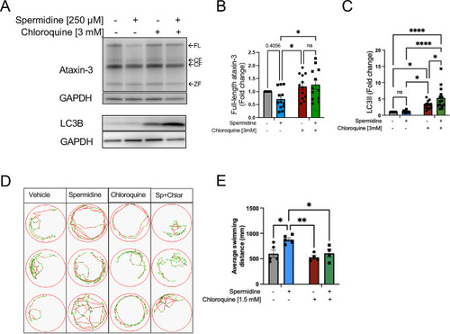
Spermidine treatment of MJD zebrafish resulted in autophagy induction and inhibition of the autophagy pathway prevented the improved swimming produced by spermidine treatment. (A) Cohorts of MJD zebrafish larvae were euthanised and processed for immunoblot analysis for ataxin-3, LC3B and GAPDH loading control. (B) Inhibition of the autophagy pathway with chloroquine resulted in a significant increase in full length ataxin-3 levels (p < 0.0459, n = 11–12). (C) Comparison of LC3II levels in lysates from zebrafish treated with chloroquine, versus those co-treated with chloroquine and spermidine revealed that spermidine treated produced a higher level of LC3II, indicative of induction of autophagy (p < 0.0167, n = 11–12). (D) Example swimming trajectories of MJD zebrafish larvae treated with vehicle, spermidine, chloroquine or spermidine + chloroquine (Sp + Chlor) during an escape response to darkness test showed that spermidine treated larvae spent more time swimming at fast speeds and less at slow speeds. Co-treatment with spermidine + chloroquine returned the swimming to more similar to the vehicle treated group. (E) Measurement of distances swum by the MJD zebrafish show that spermidine treatment significantly increased the distances swum but chloroquine co-treatment prevented the improvement produced by the spermidine treatment (p < 0.0295, n = 4–5). A two-way ANOVA was utilised for statistical analysis followed by Tukey post hoc. Data represents mean ± SEM. Data points reflected are experimental treatments of approximately 20–25 larvae

Spermidine treatment did not improve the motor impairment of CMVMJD135 mice developed motor impairment. (A) A schematic diagram illustrates that CMVMJD135 mice were treated with spermidine (3 mM) within their drinking water from 5 to 25 weeks old. Behavioural testing was conducted throughout the study and a subset of animals were euthanised at 18 weeks old and another at 25 weeks old. (B) Comparison of water intake between mice treated with spermidine and water found no significant difference (p = 0.809). (C) MJD mice had lower body weights than wild-type mice from 9 weeks old onwards (p < 0.0236), and although spermidine treatment appeared to cause decreased weights in the wild type mice, this was not a statistically significant effect. (D) Whilst mutant CMVMJD135 mice developed increased neurological scores, indicative of neurological impairment, from around 9 weeks old onwards (*p < 0.0193), treatment with spermidine did not improve this impairment (it worsened it at 7 and 12 weeks old (p < 0.0402). (E) MJD mice had decreased latencies on the rotarod compared to wild-type mice from around 9 weeks old (**p < 0.0001). Treatment with spermidine did not increase the latency before falling from an accelerating rotarod. In fact, treatment with spermidine treatment decreased the latency to fall in wild type mice compared to water treated wild type mice at 6 and 7 weeks old (***p < 0.0331). (F) MJD mice took longer to cross the balance beam than wild-type mice at 16 and 18 weeks old (**p < 0.0106). Spermidine treatment did not affect the time taken by mice to cross a balance beam, regardless of genotype. A repeated one-way ANOVA was utilised for statistical analysis followed by Tukey post hoc. Data represents mean ± SEM. Biorender.com was used to make the image in panel A

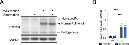
Immunoblotting of wild-type and transgenic CMVMJD135 mice confirms increased expression of full-length ataxin-3 in the cerebellum of 18-week-old MJD mice. (A) Representative western blot of 18-week-old wild-type and MJD mouse cerebellum following oral vehicle or spermidine supplementation confirming expression of full-length and endogenous ataxin-3. (B) Quantification of full-length ataxin-3 expression shows significant increase in the cerebellum of 18-week-old MJD mice compared to wild-type mice (p < 0.0001, n = 6–7). Expression of full-length ataxin-3 is not significantly altered following vehicle or spermidine treatment. A two-way ANOVA was utilised for statistical analysis followed by Tukey post hoc. Data represents means ± SEM. *** Represents p < 0.0001

Spermidine treatment did not affect presence of protein aggregates within the pons and medulla of 18-week-old CMVMJD135 mice. (A) Immunohistochemical staining for ataxin-3 in sections from the pons and medulla of wild type (WT) and CMVMJD135 (MJD) mice revealed that the MJD mice harboured ataxin-3-positive protein aggregates. Scale bars indicate 50 μm. (B) MJD mice exhibited more ataxin-3 aggregates than WT mice within the pons (p < 0.0001) and spermidine treatment had no effect on the number of ataxin-3 aggregates present. (C) MJD mice exhibited more ataxin-3 aggregates within the medulla oblongata than WT mice (p < 0.0001) and spermidine treatment had no beneficial effect on the number of ataxin-3 aggregates present, in fact greater aggregate numbers were found in the spermidine treated MJD mice compared to those treated with water (p = 0.0059). (D) More aggregates were found within the DCN region of MJD mice than WT mice (p = 0.0308), but spermidine treatment did not affect the number of aggregates present. A two-way ANOVA was utilised for statistical analysis followed by Tukey post hoc. The p-values marked on the graphs represent the Tukey test results. Data represents mean ± SEM

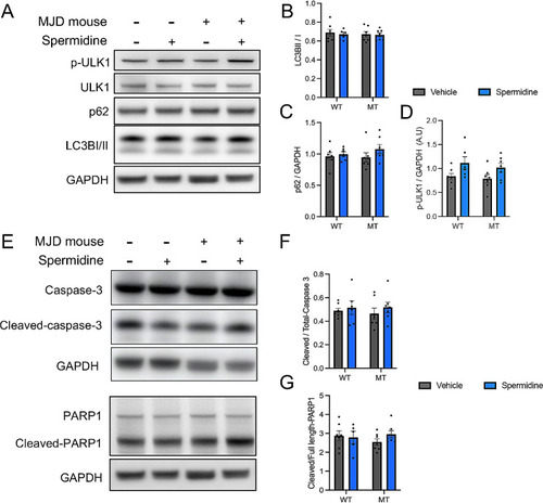
Spermidine treatment within drinking water promotes autophagy through increased phosphorylation of ULK1 in mice independent of genotype but does not induce apoptosis. (A) Representative western blot of cerebellum from wild-type and MJD mice treated with vehicle or spermidine and probed for markers of autophagy p-ULK1, ULK, p62 and LC3B. (B) Quantification of the ratio between LC3II/I showed no significant differences after spermidine treatment. (C) Quantification of p62 showed no significant differences following spermidine treatment (D) Simple main effects analysis of p-ULK1 levels demonstrated a significant increase following spermidine treatment ( p = 0.0109, n = 6–7), independent of genotype. (E) Representative western blot of wild-type and MJD mice treated with vehicle or spermidine and probed for markers of apoptosis including cleaved- and total-caspase 3 and downstream substrates cleaved- and total-PARP1. (F) Quantification of cleaved- and total-caspase 3 showed no significant differences following spermidine treatment. (G) Quantification of the ratio between cleaved- and full-length-PARP1 revealed no significant differences following spermidine treatment. A two-way ANOVA was utilised for statistical analysis followed by Tukey post hoc analysis. Data represents mean ± SEM. * Represents p < 0.05

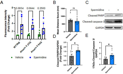
Signs of increased apoptosis in spermidine treated zebrafish larvae. (A) Non-transgenic (WTTAB) zebrafish larvae aged 2 days post fertlisation (dpf) showed increased acridine orange apoptosis staining following spermidine treatment (p = 0.0054). The increase in acridine orange staining in spermidine treated Ubb-Ataxin-3 84Q BFP zebrafish larvae was not statistically significant (p > 0.0644). (B) Comparison of the motor behaviour (total distance swum) of WT TAB zebrafish larvae treated with Vehicle or Spermidine found no difference between the groups. (C) Immunoblot analysis was performed to examine signs of apoptosis in non-transgenic zebrafish following spermidine treatment. (D) Spermidine treatment resulted in increased levels of cleaved PARP and (E) cleaved caspase 3, both signs of apoptosis (p = 0.0196 and p = 0.0255 respectively, n = 7). Statistical analysis performed were a two-way ANOVA followed by a Tukey post-hoc analysis and a paired Student t-test. Data represents mean ± SEM. Data points reflected are experimental treatments of approximately 20–25 larvae

Acknowledgments
This image is the copyrighted work of the attributed author or publisher, and ZFIN has permission only to display this image to its users. Additional permissions should be obtained from the applicable author or publisher of the image. Full text @ Mol. Brain