FIGURE SUMMARY
Title

Impaired glycine neurotransmission causes adolescent idiopathic scoliosis

Authors
Wang, X., Yue, M., Cheung, J.P.Y., Cheung, P.W.H., Fan, Y., Wu, M., Wang, X., Zhao, S., Khanshour, A.M., Rios, J.J., Chen, Z., Wang, X., Tu, W., Chan, D., Yuan, Q., Qin, D., Qiu, G., Wu, Z., Zhang, J., Ikegawa, S., Wu, N., Wise, C.A., Hu, Y., Luk, K.D.K., Song, Y.Q., Gao, B.
Source
Full text @ Journal of Clin. Invest.

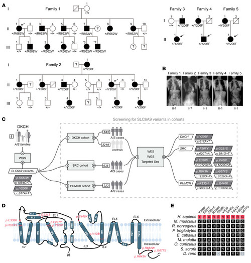
Heterozygous missense variants in SLC6A9 leading to AIS.

(A) Pedigree of 5 AIS families with dominant inheritance. Squares and circles denote male and female family members, respectively. Filled and open symbols represent affected and unaffected family members, respectively. Individuals marked with numbers indicate the family members recruited in this study. Question marks and diagonal slashes indicate unavailable and deceased members, respectively. The term +/+ denotes WT, and +/R662W or +/Y206F denotes heterozygous variant of SLC6A9. Arrows indicate the probands. (B) Spinal radiographs of the probands of family 1-5. (C) Workflow for the identification of SLC6A9 variants in the multicenter AIS cohort. The number of families and sporadic patients enrolled from each involved hospital is indicated, and the identified SLC6A9 variants are highlighted in the oblique boxes. (D) Membrane topological features of GLYT1. Positions of the identified variants are indicated in the diagram. TM, transmembrane; IL, intracellular loop; EL, extracellular loop. N and C indicate the N- and C-termini of GLYT1, respectively. (E) Evolutionary conservation of altered GLYT1 amino acids associated with AIS. Each variant is shown on the top.

Plasma glycine concentration and paraspinal muscle activity in SLC6A9 variant carriers.

(A) Plasma glycine concentration was measured in 15 AIS patients carrying SLC6A9 variants and 36 noncarrier controls (left panels). Plasma glycine concentration was measured in 6 adolescent patients carrying SLC6A9 variants and 29 adolescent controls (right panel). Boxes show the median and IQRs, with all individual data points superimposed. Orange dots represent III-6 and III-7 in family 1. Unpaired Student’s t test. **P = 0.0012; ***P = 0.00080. (B) Placing positions of bipolar electrodes. Electrodes were positioned at thoracic vertebra T9-11 in controls or at apex vertebra in AIS patients. (C) sEMG signals from healthy controls (left) and preadolescent AIS patients (right). Raw sEMG signals were collected from the paraspinal muscle at thoracic vertebra T9-11 in controls or at apex vertebra (T9-11) in patients during 5 seconds of standing in an upright posture. Blue and red represent left (L) and right (R) or concave and convex sEMG signals, respectively. (D) sEMG signals from adult AIS patient II-7, father of III-6 and III-7, in family 1. Raw sEMG signals were collected from the paraspinal muscle at apex vertebra T5-7 during 5 seconds of standing in an upright posture.

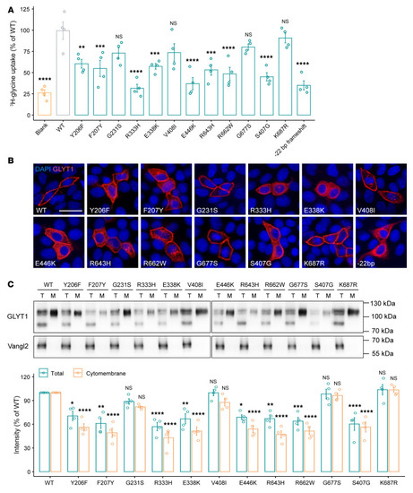
Effect of GLYT1 variants on glycine uptake and membrane presentation.

(A) Results of glycine-uptake assay for GLYT1 in HEK293T cells. Each dot represents 1 independent experiment (n = 4). Error bars represent 95% CIs. Data are represented as means ± SEM. One-way ANOVA test. **P < 0.01; ***P < 0.001; ****P < 0.0001. (B) Subcellular localization of Flag-tagged GLYT1 in MDCK cells. Signals were visualized with anti-Flag antibody (red), and nuclei were stained with DAPI (blue). Scale bar: 20 μm. (C) Western blot analysis of Flag-tagged GLYT1 in total cell lysates (T) and biotinylated membrane fractions (M) of transfected HEK293T cells. Lower and higher bands indicate underglycosylated and glycosylated GLYT1, respectively. Expression of an unrelated membrane protein, HA-tagged Vangl2, served as internal transfection control. Quantification of immunoblots of total cell extracts and cytomembrane fractions of GLYT1 variants, normalized to Vangl2 and GLYT1 WT, is shown below. Each data dot represents 1 independent experiment (n = 4). Data are represented as means ± SEM. Two-way ANOVA test. *P < 0.05; **P < 0.01; ***P < 0.001; ****P < 0.0001.

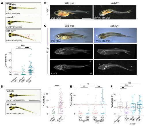
Body curvature in slc6a9 mutant zebrafish.

(A) Axial curvature of slc6a9 mutant zebrafish at 7 dpf. The severity of the curvature is measured by θ angle. (B) Spinal curvature of slc6a9 mutant zebrafish at 21 dpf. (C) Curvature phenotype and micro-CT images of WT and slc6a9m/+ zebrafish at adolescent stage (35 dpf). Images are shown in either side or dorsal view. A, anterior; P, posterior; L, left; R, right. (D) Axial curvature of WT zebrafish larvae treated with vehicle or GLYT1 inhibitor ALX 5407 (1 μM). (E) Quantification of axial curvature in WT and slc6a9m/+ zebrafish treated with vehicle (Veh) or low-dose ALX5407 (ALX, 10 nM). Only 10% of slc6a9m/+ fish showed axial curvature (θ ≥10°), whereas 10 nM ALX5407 induced axial curvature in 4.65% of WT and 34.9% of slc6a9m/+ fish. (F) Quantification of axial curvature in slc6a9m/m zebrafish with and without injection of 200 pg SLC6A9 WT or mutant (Y206F or R662W) mRNAs. Scale bars: 1 mm (A and D); 2 mm (B and C). In all charts, boxes show the median and IQRs with all individual data points superimposed. The number of analyzed fish and the penetrance of curvature (θ ≥10°) are quantified and indicated for each genotype. Unpaired Student’s t test (D) or 1-way ANOVA test (A, E, and F). **P < 0.01; ****P < 0.0001.

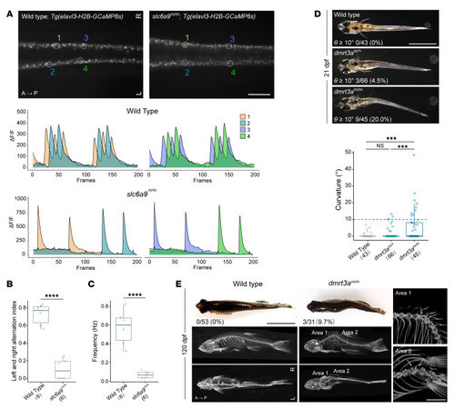
Body curvature caused by disturbance of CPG.

(A) Dorsal view fluorescent snapshots of the spinal cord of WT and slc6a9m/m zebrafish in a Tg(elavl3-H2B-GCaMP6s) background at 24 hpf. ROI is circled and numbered as 1–4. Lower panel shows quantification of fluorescence changes in the ROIs of WT and slc6a9m/m zebrafish. Each frame was taken with a 100 ms exposure and at 10 fps. GCaMP6s fluorescence intensity was defined as the ΔF/F, and ΔF/F changes within a 20-second recording time are shown. (B) Quantification of left and right alternation index in WT (n = 6) and slc6a9m/m (n = 6) zebrafish. This analysis was performed based on quantified intensities of total left- and right-side neural activities within a 1-minute recording time period. Unpaired Student’s t test. ****P < 0.0001. (C) Frequency of neural activities in WT (n = 6) and slc6a9m/m (n = 6) zebrafish. Frequency (Hz) was calculated based on left-side neural activity. Unpaired Student’s t test. ****P < 0.0001. (D) Spinal curvature of dmrt3a mutant zebrafish at 21 dpf. (E) Curvature phenotype and micro-CT images of dmrt3a mutant zebrafish at 120 dpf. Images are shown in either side or dorsal view. To detect the details of apices of curvatures, the 2 curvature regions (areas 1 and 2) of dmrt3am/m zebrafish are enlarged and oriented in different angles (right). Note that all highlighted adjacent vertebrae (arrows) are morphologically normal. Scale bars: 200 μm (A); 2 mm (D, E, right); 1 cm (E, left). Boxes show median and IQRs with all individual data points superimposed. Number of analyzed fish and the penetrance of curvature (θ ≥10°) are quantified and indicated for each genotype. Unpaired Student’s t test (B and C) or 1-way ANOVA test (D). ***P < 0.001; ****P < 0.0001.

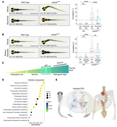
Prevention of body curvature in slc6a9 mutant zebrafish by pharmacological intervention.

(A) Representative dorsal-view images of axial phenotypes of WT and slc6a9m/m larvae treated with vehicle or strychnine (GlyR antagonist, 0.5 μM). (B) Representative dorsal-view images of axial phenotypes of WT and slc6a9m/m larvae treated with vehicle or sodium benzoate (glycine neutralizer, 0.5 ppm). Scale bars: 1 mm. In A and B, the number of analyzed fish and the penetrance of curvature are quantified and indicated for each genotype. Boxes show the median and IQRs with all individual data points superimposed. One-way ANOVA test. **P < 0.01; ****P < 0.0001. (C) Proposed glycinopathy spectrum. Abnormally high levels of glycine are associated with glycine encephalopathy, a severe neurological disease, whereas moderately elevated levels of glycine are a causal risk factor for AIS. (D) Cellular component of GO functional enrichment analysis of AIS-associated genes. GeneRatio is the ratio of genes mapped to a pathway to the total gene set. The size of the dots represents the number of genes mapped to the pathway. (E) Proposed disease mechanism of spinal curvature. In zebrafish, disruption of glyt1 causes a discoordination of left-right neural activities in the spinal cord due to aberrant glycinergic neurotransmission; deletion of dmrt3a partially impairs the development of commissural interneurons and compromises the locomotor left-right alternation; both lead to an AIS-like phenotype via the disturbance of CPGs in the spinal cord. In humans, functional impairment of GLYT1 leads to elevated glycine levels, aberrant paraspinal muscle activities, and AIS. Our findings suggest that dysfunction of the CPGs induced by either excessive glycine or developmental defects is one of the major causal factors underlying the etiology of AIS. I.N., interneurons; M.N., motoneurons.

Acknowledgments
This image is the copyrighted work of the attributed author or publisher, and ZFIN has permission only to display this image to its users. Additional permissions should be obtained from the applicable author or publisher of the image. Full text @ Journal of Clin. Invest.