GCN5L1 gene KO impaired surfactant production in MLE-12 cells. A Phospholipid secretion in WT and GCN5L1-KO MLE-12 cells. B ELISAs of secreted SPs in the culture supernatant of WT and GCN5L1-KO MLE-12 cells. C Reconstruction of GCN5L1 expression repaired phospholipid secretion in GCN5L1 mutant cells. D Reconstruction of GCN5L1 expression repaired SP-B and SP-C secretion in GCN5L1 mutant cells. The results are expressed as the mean ± SD of three independent experiments; **P < 0.01; t-test

Disruption of GCN5L1 altered the expression of Cebpα and surfactant-related genes. A Q-PCR verification of the surfactant-related DEGs in GCN5L1 mutant clones. B Q-PCR detection of Cebpα mRNA expression in WT and GCN5L1-KO MLE-12 cells. C Representative immunoblot image of Cebpα protein expression levels in WT and GCN5L1-KO MLE-12 cells. D Representative immunoblot images of Cebpα protein levels in WT cells, M2 cells, M2 cells infected with lentivirus expressing exogenous Cebpα (M2 + Cebpα), and M2 cells infected with lentivirus expressing exogenous GCN5L1 (M2 + GCN5L1). E Q-PCR assays of the RNA levels of surfactant-related genes in WT cells, M2 cells, and M2 cells infected with lentivirus expressing exogenous Cebpα (M2 + Cebpα), and M2 cells infected with lentivirus expressing exogenous GCN5L1 (M2 + GCN5L1). The results are expressed as the mean ± SD of three independent experiments; ns, not significant;*P < 0.05, **P < 0.01; t-test

Disruption of GCN5L1 altered the activity of the ROS–ERK–FOXO1 axis in MLE-12 cells. A Q-PCR detection of Foxo1 mRNA expression in WT, GCN5L1-KO MLE-12 cells. B Representative immunoblot image of Foxo1 protein expression levels in WT, GCN5L1-KO MLE-12 cells and M2 cells infected with lentivirus expressing exogenous GCN5L1 (M2 + GCN5L1). C Representative immunoblot images of ERK and p-ERK protein expression levels in WT, GCN5L1-KO MLE-12 cells and M2 cells infected with lentivirus expressing exogenous GCN5L1 (M2 + GCN5L1). D Cellular ROS levels of WT, GCN5L1-KO MLE-12 cells, and M2 cells infected with lentivirus expressing exogenous GCN5L1 (M2 + GCN5L1). Cells were incubated with CM-H2DCFDA and measured by FACS; mean fluorescence intensity was normalized to WT group. E Representative immunoblot images of ERK, p-ERK, Foxo1, and Cebpα protein expression levels in WT cells, M2 cells treated with DPI (M2 + DPI) or SCH772984 (M2 + SCH772984), and M2 cells infected with lentivirus expressing exogenous Foxo1 (M2 + Foxo1). F Q-PCR assays of the RNA levels of surfactant-related genes in WT cells, M2 cells treated with DPI (M2 + DPI) or SCH772984 (M2 + SCH772984), and M2 cells infected with lentivirus expressing exogenous Foxo1 (M2 + Foxo1). The results are expressed as the mean ± SD of three independent experiments; ns, not significant; *P < 0.05,**P < 0.01; t-test

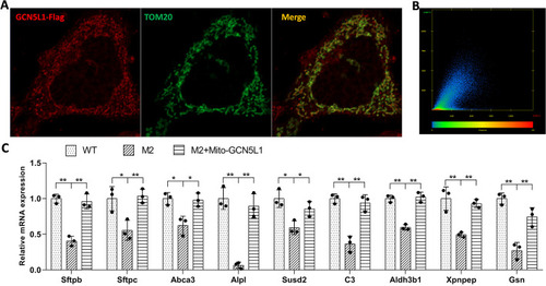
Mitochondria-localized GCN5L1 is responsible for the regulation of surfactant-related genes. A Colocalization of GCN5L1–FLAG with TOM20 in MLE-12 cells. Cells were cotransfected with TOM20- and GCN5L1–FLAG-expressing plasmids before staining with anti-TOM20 and anti-FLAG antibodies. B The colocalization scatter plot of anti-TOM20 and anti-GCN5L1–FLAG signals. Pearson correlation coefficient is 0.78, Manders’ overlap coefficient is 0.82. C Q-PCR assays of the RNA levels of surfactant related genes in WT cells, M2 cells, and M2 cells infected with lentivirus expressing mito-GCN5L1 (M2 + mito-GCN5L1).The results are expressed as the mean ± SD of three independent experiments; *P < 0.05, **P < 0.01; t-test

Impairment of LB positioning/trafficking in GCN5L1-KO cells. A Lysotracker Red staining of WT, GCN5L1-KO clones (M2 and M29), and GCN5L1 reconstructed mutant cells (M2 + GCN5L1). B WT and M2 cells were metabolically labeled with BODIPY phosphatidylcholine. C WT and M2 cells were immunostained with Lamp1 antibody. D–F WT and M2 cells were transfected with h-ABCA3–EGFP or ABCA3–mCherry before staining with Lysotracker Red (D), BODIPY phosphatidylcholine (E), and Lamp1 antibody (F)

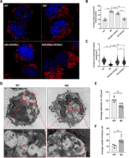
Abnormal size and number of LB-like organelles in GCN5L1-KO cells. A Representative images showing Lysotracker-stained organelles in WT, M2, M2 + GCN5L1, and M2 + mito-GCN5L1 cells. B, C The numbers and size (area) in WT, M2, M2 + GCN5L1, and M2 + mito-GCN5L1 cells; **P < 0.01 (Kolmogorov–Smirnov test). D Transmission electron micrographs of WT and M2 cells. Many LB-like organelles are present within the cytoplasm; arrows point to lamellar-like materials in the organelles. E, F The diameters and numbers of LBs in the TEM images were measured and counted with Image J. The results are expressed as the mean ± SD of four independently measured data; *P < 0.05; t-test

Localization of GCN5L1 in LBs. Cells were transfected with GCN5L1–FLAG-expressing plasmid and stained with anti-Lamp1 and anti-FLAG antibodies

Accumulation of SP proteins in GCN5L1-KO cells. A Immunoblot image of WT, M2, and GCN5L1 reconstructed M2 + GCN5L1 cells with an SP-B antibody (Santa Cruz). B Immunoblot image of WT, M2, and GCN5L1 reconstructed M2 + GCN5L1 cells with another SP-B antibody (Abcam). C WT and M2 cells were cotransfected with h-ABCA3–mCherry and m-Sftpb expressing plasmids and immunostained with an SP-B antibody (Abcam). D WT and M2 cells were cotransfected with h-ABCA3–EGFP- and m-Sftpc-expressing plasmids and immunostained with an SP-C antibody (Seven Hills Bioreagents). **P < 0.01; t-test

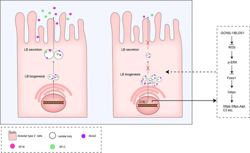
A hypothetical illustration of GCN5L1 functions in the regulation of pulmonary surfactant production

Acknowledgments
This image is the copyrighted work of the attributed author or publisher, and ZFIN has permission only to display this image to its users. Additional permissions should be obtained from the applicable author or publisher of the image. Full text @ Cell Mol. Biol. Lett.