HSPC formation is impaired in alas2- and alad-deficient embryos. (A) Screening strategy of HSPC development under different primitive RBC-deficient conditions. (B) Bright-field images of blood flow in alas2−/−, alad−/− and their siblings at 36 hpf. Blood flow in heart regions is denoted by arrowheads. (C) Cumulative results of blood color phenotype in B. Number of embryos with different blood color are noted in each column. (D,E) Relative mRNA level of runx1 (D) and cmyb (E) in the dissected trunk regions of alas2−/−, alad−/− and their sibling at 36 hpf examined by qRT-PCR. n=3 experimental replicates. (F,G) Expression of runx1 and cmyb in alas2−/−, alad−/− and their sibling at 36 hpf examined by WISH. The AGM regions for marker gene-positive cell counting are denoted by red arrowheads. (H,I) Quantification of the runx1- and cmyb-positive HSPCs in F and G. n=3 experimental replicates. (J) Confocal imaging shows the kdrl+/cmyb+ HSPCs in AGM regions of alas2−/−, alad−/− and their siblings at 36 hpf. The kdrl+/cmyb+ HSPCs in the AGM region are denoted by white arrowheads. (K) Quantification of the indicated cells in confocal imaging. n=3 experimental replicates. Number of samples are indicated. Data are mean±s.d. **P<0.01; ***P<0.001 [Mann–Whitney non-parametric U-test (D,E); one-way ANOVA, Tukey's multiple comparisons (H,I,K)]. n.s., not significant. Scale bars: 200 μm (B); 100 μm (F,G,J).

Heme-deficient primitive RBCs display iron accumulation. (A) Workflow of iron distribution assay of body and corresponding yolk regions by ICP-MS or iron colorimetric assay kit, respectively, in alas2−/−, alad−/− and their siblings at 36 hpf. (B-D) Quantification of the total iron level of dissected body (B) and yolk (C,D). The body and yolk sac iron content are quantified as weight (mg/kg) and volume (mg/l or μmol/mg protein), respectively. n=4 experimental replicates. (E) Representative bright-field images of DAB-enhanced iron staining in alas2−/−, alad−/− and their siblings at 36 hpf. Right column shows magnification of areas in red box showing iron enriched RBCs in caudal hematopoietic tissue (CHT) circulation. (F) Quantification of the iron staining-positive RBCs in E. n=3 experimental replicates. Number of samples are indicated. Data are mean±s.d. ***P<0.001 (one-way ANOVA, Tukey's multiple comparisons). n.s., not significant. Scale bars: 100 μm.

Blood IOL phenotype is caused by heme-deficient RBCs via Slc40a1. (A) Workflow of blood iron measurement in control, alas2−/− and alad−/− at 36 hpf by ICP-MS or iron colorimetric assay kit. (B,C) Quantification of the blood iron content by ICP-MS (B) and iron colorimetric assay kit (C). n=5 experimental replicates. (D) Schematic of bulk RNA-seq of flow cytometry-sorted gata1+ RBCs in alas2 and alad morphants at 36 hpf on Illumina NovaSeq 6000 platform. (E,F) GO analysis showing the enrichment of upregulated (E) and downregulated (F) overlapping terms in heme-deficient RBCs of alas2 and alad morphants. (G) Heatmap analysis of selected overlapping DEGs in iron transportation-related GO terms. The iron exporter Slc40a1 is highlighted with red dashed rectangle. (H) The expression of slc40a1 is detected by WISH in control, alas2−/− and alad−/− at 36 hpf. The RBC-enriched yolk regions are denoted by red arrowheads. (I) Statistical analysis of the relative slc40a1 expression in H by ImageJ. n (embryos)=10. (J) Relative mRNA level of slc40a1 in the flow cytometric-sorted RBCs (gata1+) of control, alas2−/− and alad−/− at 36 hpf examined by qRT-PCR. n=3 experimental replicates. (K) Expression of HSPC marker gene runx1 in control, alas2−/− and alad−/− with or without VIT2763 treatment at 36 hpf examined by WISH. The AGM regions for marker gene-positive cell counting are denoted by red arrowheads. (L) Quantification of the runx1-positive HSPCs in K. n=3 experimental replicates. Number of samples are indicated. Data are mean±s.d. ***P<0.001 (one-way ANOVA, Tukey's multiple comparisons). n.s., not significant. Scale bars: 250 μm (H); 100 μm (K).

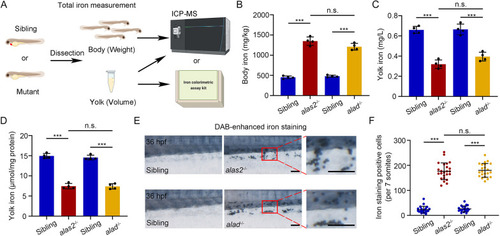
Iron-dependent HSPC ferroptosis in alas2−/− and alad−/−. (A) Transmission electron microscopy (TEM) view of a longitudinal section through the artery and vein in control, alas2−/− and alad−/− AGM region at 36 hpf. (B) TEM view of HSPC subcellular structures. From top to bottom panels are mitochondria and membrane. Arrowheads in black denote mitochondria, green denote plasma membrane and blue denote double-membrane vesicle. (C-E) Quantification of mitochondria area, mitochondria cristae area and cristae number in HSPCs of control, alas2−/− and alad−/−. Box plots show median values (middle bars) and first to third interquartile ranges (boxes); whiskers indicate 1.5 times the interquartile ranges (IQR); individual points from the 10 to 90 percentile are plotted and shown. n (mitochondria)=96. (F) Quantification of membrane-disrupted HSPCs in control, alas2−/− and alad−/−. n (HSPCs)=12. (G,H) Western-blotting detection of the protein level of Fth1, Gpx4 and Slc7a11 in flow cytometric-sorted HSPCs of control, alas2−/− and alad−/− at 36 hpf, respectively (30,000 HSPCs were sorted in each group). (I) Quantification of protein levels in H. Protein levels were analyzed using 8-bit-gray analysis (Gel-Pro analyzer). n=3 experimental replicates. (J) Schematic workflow for the ferrous iron assessment in HSPCs (cmyb+) of alas2−/− and alad−/− with fluorescent iron probes (Fe2+ biotracker dye, FeRhoNox-1 and FerroOrange) at 36 hpf. (K) Representative flow cytometric histogram of the Fe2+ level in sorted HSPCs of control, alas2−/− and alad−/− at 36 hpf measured by Fe2+ biotracker dye. (L) Quantification of mean fluorescence intensity (MFI) of labile Fe2+ level in K. (M) Representative fluorescence images show the co-localization of cmyb:GFP+ and FeRhoNox-1+ cells in alas2−/− and alad−/− at 36 hpf. The cmyb+/FeRhoNox-1+ cells are denoted by white arrowheads. (N) Flow cytometric analysis of the percentage of cmyb+/FeRhoNox-1+ cells in control, alas2−/− and alad−/− at 36 hpf. (O) Statistical analysis of the percentage of FeRhoNox-1+ HSPCs in N. n=3 experimental replicates. (P) Quantification of MFI of Fe2+ level in N. n=3 experimental replicates. (Q) Expression of HSPC marker runx1 in control, alas2−/− and alad−/− with or without DFO treatment (100 μM) at 36 hpf examined by WISH. The AGM regions for marker gene-positive cell counting are denoted by red arrowheads. (R) Quantification of the runx1-positive HSPCs in Q. n=3 experimental replicates. (S) Confocal imaging shows the kdrl+/cmyb+ HSPCs in control, alas2−/− and alad−/− with or without DFO treatment at 36 hpf. (T) Quantification of the HSPCs in S. n=3 experimental replicates. (U) Expression of HSPC marker runx1 in control, alas2−/− and alad−/− with or without Ferrostatin-1 (Fer-1) treatment (10 μM) at 36 hpf examined by WISH. The AGM regions for marker gene-positive cell counting are denoted by red arrowheads. (V) Quantification of the runx1-positive HSPCs in U. n=3 experimental replicates. (W) Confocal imaging shows the kdrl+/cmyb+ HSPCs in control, alas2−/− and alad−/− with or without Fer-1 treatment at 36 hpf. (X) Quantification of the HSPCs in W. n=3 experimental replicates. Number of samples are indicated. Data are mean±s.d. *P<0.05, **P<0.01, ***P<0.001 [one-way ANOVA, Tukey's multiple comparisons (C-F,L,O,P,R,T,V,X); two-way ANOVA, Sidak's multiple comparisons (I)]. n.s., not significant. Scale bars: 10 μm (A); 0.5 μm (B); 50 μm (M); 100 μm (Q,S,U,W).

The HSPC ROS level is elevated in alas2 and alad mutants. (A) Schematic of biochemical assay of oxidative stress biomarkers (ROS, CAT, SOD, GSH and MDA) in zebrafish dissected trunk regions at 36 hpf. (B) Confocal imaging showing the co-localization of kdrl:GFP+ and MitoSOX+ cells in the VDA of control, alas2−/− and alad−/− at 36 hpf. The dorsal aorta (DA) regions are denoted by white dashed lines, and the kdrl+/MitoSOX+ cells are denoted by white arrowheads. (C) Quantification of kdrl+/MitoSOX+ cells in B. n=3 experimental replicates. (D) Quantification of mean fluorescence intensity (MFI) of mitochondrial ROS level in HSPCs of control, alas2−/− and alad−/− measured by MitoSOX staining. n=3 experimental replicates. (E) Expression of the HSPC marker runx1 in control, alas2−/− and alad−/− with or without NAC treatment at 36 hpf examined by WISH. The AGM regions for marker gene-positive cell counting are denoted by red arrowheads. (F) Quantification of the runx1-positive HSPCs in E. n=3 experimental replicates. (G) Expression of the HSPC marker runx1 in control, alas2−/− and alad−/− with or without mitoTempo treatment at 36 hpf examined by WISH. The AGM regions for marker gene-positive cell counting are denoted by red arrowheads. (H) Quantification of the runx1-positive HSPCs in G. n=3 experimental replicates. (I) Confocal imaging shows the kdrl+/cmyb+ HSPCs in control, alas2−/− and alad−/− with or without mitoTempo treatment at 36 hpf. (J) Quantification of the HSPCs in I.; n=3 experimental replicates. Number of samples are indicated. Data are mean±s.d. ***P<0.001 (one-way ANOVA, Tukey's multiple comparisons in C,D,F,H,J). n.s., not significant. Scale bars: 100 μm.

Iron-mediated AA oxidation leads to overproduction of 5S- and 12S-related ferroptotic stimulus. (A) Schematic workflow for the metabolomics analyses using the dissected trunk region of control, alas2- and alad-morphants at 36 hpf. (B) Pie-chart of the proportion of each lipid species in total oxidative lipids. AA, arachidonic acid; ALA, α-linoleic acid; DGLA, dihomo-γ- linoleic acid; DHA, docosahexaenoic acid; EPA, eicosapentaenoic acid; LA, linoleic acid. (C) Heatmaps of differentially regulated oxidative lipids in alas2 and alad morphants. 5S- and 12S-related oxidative lipids are denoted by red arrowheads. (D) Schematic workflow for the MALDI-MSI experiment using control, alas2−/− and alad−/− at 36 hpf. Cross-cryosections of AGM region are collected (denoted by red dashed rectangle). (E) High resolution MALDI-MS images of succinyl-CoA, AA and 1-arachidonoyl glycerophosphoinositol in control, alas2−/− and alad−/− at 36 hpf. Left panel is the navigator for cross-section. Relative abundance (mass spectrum peak intensity) of each metabolite is labeled as per the color bars. (F) Statistical analyses of the relative abundance of succinyl-CoA, AA and 1-arachidonoyl glycerophosphoinositol in the VDA regions in H. n=2-4 experimental replicates. (G) Relative MDA content in control, alas2−/− and alad−/− with or without PRGL493 treatment at 36 hpf. PRGL493 treatment from 26 to 36 hpf. n=3 experimental replicates. (H) Expression of the HSPC marker runx1 in control, alas2−/− and alad−/− with or without PRGL493 treatment at 36 hpf examined by WISH. The AGM regions for marker gene-positive cell counting are denoted by red arrowheads. (I) Quantification of the runx1-positive HSPCs in H. n=3 experimental replicates. Number of samples are indicated. Data are mean±s.d. *P<0.05, **P<0.01, ***P<0.001 (one-way ANOVA, Tukey's multiple comparisons). n.s., not significant. Scale bars: 100 μm.

HSPCs derived from alas2- and alad-deficient embryos show low erythroid lineage reconstitution efficiency. (A) Schematic workflow for the primary and secondary HSPC transplantation with sorted ubi:dsRed+/CD41:GFPlow HSPCs in alas2 and alad morphants at 5 dpf. (B) Flow cytometric analysis of engrafted ubi-dsRed+ cells in the recipient kidney marrow (KM) at 45 dpt. Number of recipients: n (control HSPCs>irradiated WT)=8, n (alas2-morphant HSPCs>irradiated WT)=6, n (alad-morphant HSPCs>irradiated WT)=7. (C) Statistical analysis of the engraftment efficiency in B; the total numbers of recipients are indicated. (D) Flow cytometric analysis of different blood cell types in recipient kidney marrows. Each circle and percentile represent different cell lineages (I, erythrocytes; II, myelocytes; III, lymphocytes; IV, progenitors). (E) Quantification of different blood lineages in the kidney marrows based on D. Total cell numbers in whole kidney marrows are indicated. (F) Statistical analysis of the number of RBCs in recipient peripheral blood. The total numbers of recipients are indicated. (G) Statistical analysis of the engraftment efficiency of control, alas2 and alad morphants after secondary transplantation. Number of recipients: n (control HSPCs>irradiated WT)=7, n (alas2-morphant HSPCs>irradiated WT)=7, n (alad-morphant HSPCs>irradiated WT)=5. Total numbers of recipients are indicated. (H) Quantification of different blood lineages in the kidney marrows based on G. Total numbers of recipients are indicated. Number of samples are indicated. Data are mean±s.d. **P<0.01, ***P<0.001 [one-way ANOVA, Tukey's multiple comparisons (C,F,G); two-way ANOVA, Sidak's multiple comparisons (E,H)]. n.s., not significant.

Acknowledgments
This image is the copyrighted work of the attributed author or publisher, and ZFIN has permission only to display this image to its users. Additional permissions should be obtained from the applicable author or publisher of the image. Full text @ Development