FIGURE SUMMARY
Title

Characterization of the Interrenal Gland and Sexual Traits Development in cyp17a2-Deficient Zebrafish

Authors
Shi, S., Shu, T., Li, X., Lou, Q., Jin, X., He, J., Yin, Z., Zhai, G.
Source
Full text @ Front Endocrinol (Lausanne)

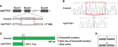
Deletion of cyp17a2 in zebrafish by CRISPR/Cas9. (A) The gRNA target site of cyp17a2 in the second exon. cyp17a2-null zebrafish with 7 base pairs deletion was obtained. (B) The representative chromatograms of the DNA sequences of control fish and cyp17a2-/- zebrafish, as evidenced by DNA sequencing of the PCR amplified DNA fragments using the genome DNA or cDNA as the template. The red squares in panel A and B indicate the 7 base pairs which have been deleted in cyp17a2-/- fish. (C) The predicted Cyp17a2 protein and mutant protein of cyp17a2-/- fish. The peptides of mutant Cyp17a2 identical to control Cyp17a2 protein are shown in green, the frameshift mutation is shown in gray. The premature stop codon is indicated with purple asterisk in panel (C) aa, amino acid. The amino acids before and after the start site of frameshift mutation in cyp17a2-/- fish and the corresponding area in the control fish is highlighted in dotted square and the detailed information is shown in panel (D). (D) The detailed amino acid sequence of the dotted square in panel (C).

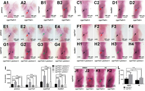
cyp17a2-deficiency caused pituitary pomca upregulation and interrenal gland enlargement in zebrafish larvae. WISH analyses with probes of pomca(A1, A2), cyp11a1(B1, B2), cyp21a2(C1, C2) and hsd3b1(D1, D2) in cyp17a2+/+ and cyp17a2-/- fish at 5 dpf. WISH analyses with probes of pomca(E1–E4), cyp11a1(F1–F4), cyp21a2(G1–G4), and hsd3b1(H1–H4) in fish at 5 dpf. (E1, F1, G1, H1)cyp17a2+/+;pomca+/+ fish. (E2, F2, G2, H2)cyp17a2-/-;pomca+/+ fish. (E3, F3, G3, H3)cyp17a2+/+;pomca-/- fish. (E4, F4, G4, H4)cyp17a2-/-;pomca-/- fish. Black, red, and green arrows indicate normal, enlarged, and decreased interrenal gland, respectively. (I) Bar chart represents the quantification and statistical analyses of WISH signals in pituitary and interrenal gland of fish at 5 dpf. (J1, J2, K1, K2) WISH analysis using the probe of cyp21a2 in cyp17a2+/+ and cyp17a2-/- fish administrated with DMSO or HC from 1 dpf to 5 dpf. (J1, J2) DMSO. (K1, K2) HC. (L) Bar chart represents the quantification and statistical analyses of WISH signals in interrenal gland of fish administrated with DMSO or HC. HC, hydrocortisone. DMSO, dimethyl sulfoxide. n.s., no significant difference. *P < 0.05. **P < 0.01. ***P < 0.001. ****P < 0.0001.

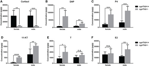
The steroid measurements. Whole-body levels of cortisol (A), DHP (B), P4 (C), 11-KT (D), T (E), and E2 (F) in cyp17a2+/+ and cyp17a2-/- zebrafish at 3 mpf (n = 6). n.s., no significant difference. *P < 0.05. **P < 0.01. ***P < 0.001. ****P < 0.0001.

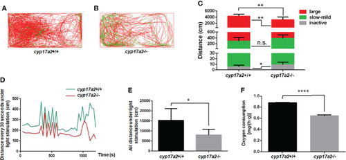
cyp17a2-deficiency resulted in impaired locomotion activity and stress response to light in zebrafish. (A, B) The locomotor trajectories of cyp17a2+/+ and cyp17a2-/- adult fish within 5 minutes (n = 10). (C) The distance fish covered at large, slow-mild, and inactive speed. The locomotor trajectories and distance covered at large (> 5 cm/sec), slow-mild (2-5 cm/sec) and inactive (< 2 cm/sec) speed in panel A-C are highlighted in red, green, and grey, respectively. (D) The movement distance curve analyzed in every 30 seconds under the light-stimulated stress within 20 minutes (n = 5). (E) All distance under the light-stimulated stress within 20 minutes (n = 5). (F) The oxygen consumption of cyp17a2+/+ and cyp17a2-/- fish after 2 days starvation (n = 3/group, 3 groups per genotype). n.s., no significant difference. *P < 0.05. **P < 0.01. ****P < 0.0001.

The analyses of primary and secondary sex characters and fertility assessment. (A–D) Gross appearance of cyp17a2+/+ male, cyp17a2-/- male, cyp17a2+/+ female, cyp17a2-/- female (n = 10). (E–H) The genital papilla. (I–L) The pectoral fin. (M–P) The anatomical observations. (Q–T) The histological analysis of gonads. Black and green arrow indicates normal and diminished genital papilla, respectively. (U) GSI of cyp17a2+/+ fish and cyp17a2-/- fish at 3 mpf (n > 10). (V) The fertility analysis of cyp17a2-/- males and cyp17a2-/- females mated with WT females and males, respectively (n = 10). (W) The statistical analyses of ovulated eggs of cyp17a2+/+, cyp17a2+/- and cyp17a2-/- females mated with WT males (n = 10). (X) The statistical analyses of fertilization ratio of the ovulated eggs from WT females when mated with cyp17a2+/+ males and cyp17a2-/- males, respectively (n = 10). n.s., no significant difference. *P < 0.05. **P < 0.01. ***P < 0.001.

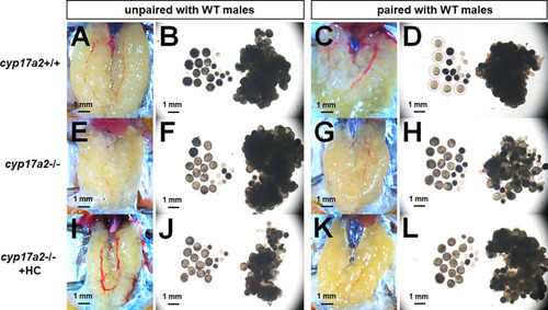
The over-activated oocytes in cyp17a2-/- females. (A–D) Under visual microscopy, the oocytes from control females unpaired and paired with WT males were not transparent and transparent, respectively (n = 5). (E–H) The oocytes from cyp17a2-/- females unpaired and paired with WT males were both transparent (n = 5). (I–L) The 20 nM hydrocortisone treatment in cyp17a2-/- females did not promotes oocyte maturation as observed under visual microscopy (n = 5).

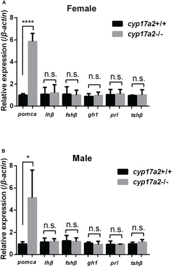
Expression profiles of pituitary hormones. Expressions of pomca, lhβ, fshβ, gh1, prl and tshβ in cyp17a2+/+ and cyp17a2-/- zebrafish at 3 mpf were analyzed with qPCR. All mRNA levels were calculated as the fold expression relative to the housekeeping gene β-actin. (A) Control females and cyp17a2-/- females. (B) Control males and cyp17a2-/- males. n.s., no significant difference. *P < 0.05. ****P < 0.0001.

Acknowledgments
This image is the copyrighted work of the attributed author or publisher, and ZFIN has permission only to display this image to its users. Additional permissions should be obtained from the applicable author or publisher of the image. Full text @ Front Endocrinol (Lausanne)