FIGURE SUMMARY
Title

Hnrnpul1 controls transcription, splicing, and modulates skeletal and limb development in vivo

Authors
Blackwell, D.L., Fraser, S.D., Caluseriu, O., Vivori, C., Tyndall, A.V., Lamont, R.E., Parboosingh, J.S., Innes, A.M., Bernier, F.P., Childs, S.J.
Source
Full text @ G3 (Bethesda)

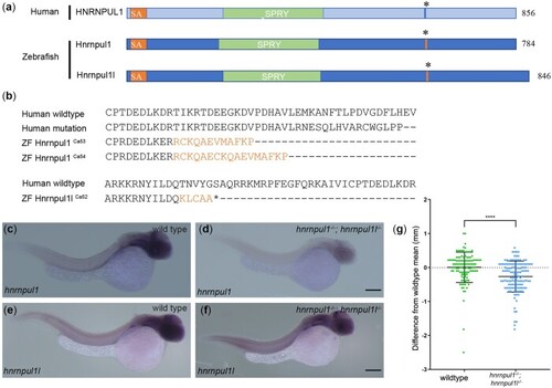
Mutation of human HNRNPUL1 and zebrafish hnrnpul1 and hnrnpul1l. a) Schematic showing domains of human HNRNPUL1 and zebrafish Hnrnpul1 and Hnrnpul1l proteins. The mutation location in human HNRNPUL1 and equivalent sequence in zebrafish is marked by *. b) Comparisons of the locations of human HNRNPUL1 wild type, human mutant, and zebrafish hnrnpul1ca53 and ca54 protein sequences showing frameshifted sequence (lighter font) following the mutation. Zebrafish hnrnpul1l wild type is compared to zebrafish hnrnpul1l in the exon 11 region where splicing to exon 13 creates a frameshift and stop codon after the new splice junction. c–f) WISH staining for hnrnpul1 (c, d) and hnrnpul1l (e, f), at 24 hpf reveals expression that is not spatially restricted in wild types (c, e), but is reduced in maternal zygotic (MZ) hnrnpul1/1l mutants (d, f). g) Quantification of SL normalized to the mean wild-type SL of 16 dpf larvae. Wild type n= 115, hnrnpul1/1l MZ mutant n= 127. ****P < 0.0001, determined by Student’s t-test. Scale bars = 200 µm.

Loss of hnrnpul1l and hnrnpul1 leads to differential splicing. a) Schematic showing the processing of hnrnpul1l to form standard transcript and AS of exon 12 of the hnrnpul1l gene as a result of CRISPR-Cas9 targeted mutagenesis. b) PSI for exon 12 of hnrnpul1l in wild type and hnrnpul1/1l mutant embryos at 3 dpf. c) Change in PSI of all exon skipping events in hnrnpul1/1l mutant embryos compared to wild types. d) Detailed view of PSI of genes associated with phenotypes, points represent each biological replicate. e) Change in percent intron retention (PIR) of all intron retention events in hnrnpul1/1l mutant embryos compared to wild types. f) Detailed view of PIR of genes associated with phenotypes, points represent each biological replicate. Details of affected exon/intron are given in Supplementary Table 1. g) Volcano plot showing all differentially expressed genes. Gray points = 1>Log2 FC>-1 or P > 0.05, orange points = 1<Log2 FC<-1 and P ≤ 0.05, green points = 1<Log2 FC<-1 and P ≤ 0.01. h) qPCR validation hnrnpul1 and hnrnpul1l expression in hnrnpul1/1l mutants at 3 dpf compared to wild types. **P ≤ 0.01, ****P ≤ 0.0001, determined by Student’s t-test.

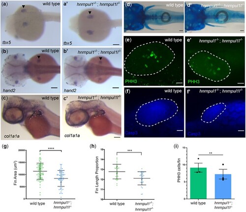
Loss of hnrnpul1 and hnrnpul1l does not affect fin specification, but leads to decreased fin growth in embryos and larvae. a–c) mRNA expression of fin specification markers tbx5 (a, a’) at 24 hpf, and hand2 (b, b’) at 48 hpf in wild type (a, b) and hnrnpul1/1l mutant (a’, b’) embryos. c, c’) mRNA expression of col1a1a in wild type and hnrnpul1/1l mutant embryos at 48 hpf. Dashed line shows outside edge of staining where fin size was measured. d, d’) Alcian blue cartilage staining of wild type and hnrnpul1/1l mutant fish at 16 dpf. e, e’) PHH3 staining of wild type and hnrnpul1/1l mutant fins at 48 hpf. f, f’) Activated Caspase 3 (Casp3) immunostaining in wild type and hnrnpul1/1l mutant fins at 48 hpf. Dotted lines show the fin boundary. g) Quantification of fin area in wild type and hnrnpul1/1l mutant col1a1a stained embryos at 48 hpf. Wild type n = 79, hnrnpul1/1ln= 68, from 2 trials. h) Quantification of fin length at 16 dpf as a percentage of body length. Wild type n = 28, hnrnpul1/1ln = 27, from 2 trials. i) Quantification of proliferation via PHH3 immunostaining in wild type and hnrnpul1/1l mutant fins at 48 hpf, n = 30 fins, from 3 trials. **P ≤ 0.01, ***P ≤ 0.001, ****P ≤ 0.0001, ns = P > 0.05, determined using Student’s t-test. Scale bars: a–d’ = 100 µm, e–f’ = 20 µm.

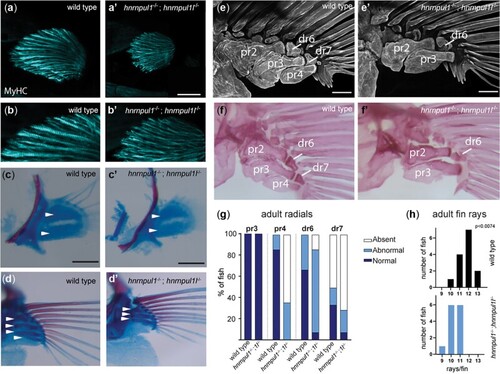
Musculoskeletal defects in hnrnpul1/1l mutant fins. a, b) Fin muscle, as highlighted by MF20 staining for myosin heavy chain (MyHC) in wild type (a, b) and hnrnpul1/1l mutant (a’, b’) fins at 72 hpf. Scale bar = 50 μm. c–f) Alcian blue (cartilage) and alarizin red (bone) staining. c, c’) At early stages (40 dpf, SL = 6.2–6.7 mm) 2 CSZs are present in both wild type and hnrnpul1/1l mutant fins (arrowheads, scale bars = 50 μm). d, d’) At larval stages (40 dpf, SL = 11 mm) there are 4 proximal radial precursors in wild-type fins, but 3 proximal radial precursors in hnrnpul1/1l mutant fins (arrowheads; scale bars = 100 μm). e, f’) In adult fish (3-month old), alizarin red staining fluorescent imaged (e, e’) or brightfield imaged (f, f’) shows a missing proximal radial (pr4) and distal radial 7 (dr7) in a hnrnpul1/1l mutant. Scale bars are 200 µm. g) Quantification of the frequency of missing proximal and distal radials in adults (n = 12 wild type, 14 hnrnpul1/1l mutants). h) Quantification of the frequency of fins with the indicated number of rays per fin (n = 14 wild type, 13 hnrnpul1/1l mutant, P < 0.074, chi-squared test).

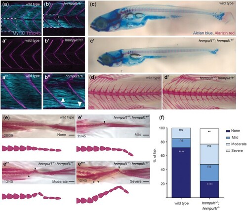
Loss of hnrnpul1l and hnrnpul1 leads to caudal scoliosis in adult Zebrafish. a, b) Muscle (MyHC, blue) and tendon (Tbsp4b, magenta) staining of 72 hpf wild type (a, a’’) and hnrnpul1/1l mutant (b, b’’) embryos, arrowheads highlight less organized muscle fibers in hnrnpul1/1l mutants. c, c’) Alcian blue cartilage and alarizin red bone staining of 16 dpf wild type and hnrnpul1/1l. d, d’) Alizarin red bone staining of 16-week-old adult wild type (d) and hnrnpul1/1l mutant (d’) fish. e, e’’’) Alizarin red bone staining of 16-week-old adult wild type and hnrnpul1/1l mutants graded by scoliosis in the caudal (tail) region (arrowheads) as none (e), mild (e’), moderate (e’’), and severe (e’’’). Schematics show spine curvature from the images above. f) Quantification of the proportion of fish with none, mild, moderate, or severe scoliosis. Wild type n = 39, hnrnpul1/1l mutant n = 45, from 3 trials. **P ≤ 0.01, ****P ≤ 0.0001 determined by Fisher’s exact test. Scale bar = 1000 µm (e, e’’’).

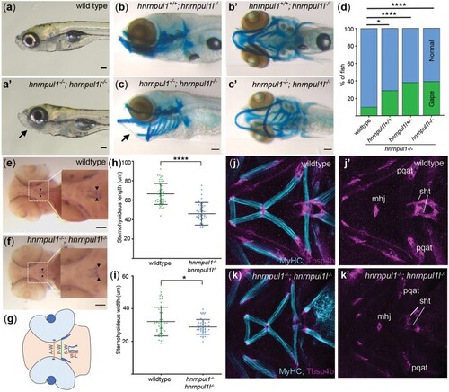
hnrnpul1/1l mutants show a shortened sternohyoideus tendon. a, a’) Images of live 8 dpf wild type and hnrnpul1/1l mutant larvae. b–c’) Lateral (b, c), and ventral (b’, c’) Alcian blue staining at 8 dpf in normal hnrnpul1l−/− mutant (b, b’) and a gaping-jaw phenotype in hnrnpul1/1l mutant (arrows) (c, c’). d) Quantification of the proportion of fish showing a gaping-jaw phenotype. Wild type n = 283, hnrnpul1l−/−; hnrnpul+/+n = 24, hnrnpul1l−/−; hnrnpul1+/−n = 64, hnrnpul1−/−hnrnpul1l−/− n = 84 from 5 trials. e, f) WISH staining for scleraxis (scxa) in the sternohyoideus tendon (box, arrow heads) in wild type and hnrnpul1/1l mutant embryos at 72 hpf. g) Schematic showing craniofacial tendons and location of tendon measurements. A-W = width between adductor mandibulae tendons, P-W = width between Palatoquadrate tendons, S-W= width between sternohyoideus tendons, S-L = sternohyoideus length. h, i) Quantification of the length (h) and width (i) between the sternohyoideus tendons in wild type (n = 51) and hnrnpul1/1l mutant (n = 50) embryos, from 3 trials. j–k’) Muscle (MyHC, blue) and tendon (Thbs4b, magenta) staining of the craniofacial region of 72 hpf wild type (j, j’) and hnrnpul1/1l mutant (k, k’). *P ≤ 0.05, ****P ≤ 0.0001 determined by Fisher’s exact test (d) and Student’s t-test (h, j) Scale bars = 100 µm. mhj, mandibulohyoid junction; pqat, palatoquadrate adductor tendon; sht, sternohyoideus tendon.

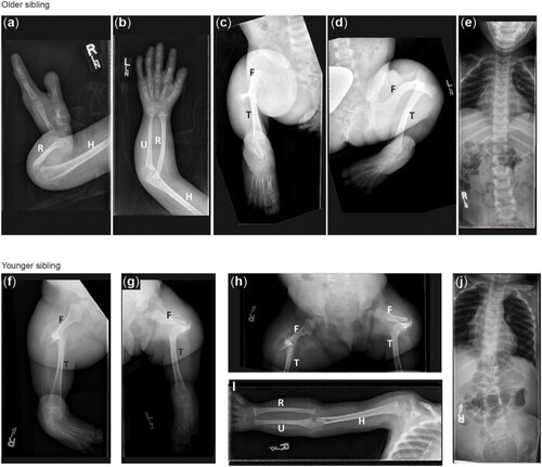
Radiographic features of siblings with a VUS in HNRNPUL1. a–e) X-ray images of older sibling. Right arm showing short humerus and absent ulna with 2 fixed in extension digits of the right hand (a). Left arm showing short humerus and normal upper arm (b). Right (c) and Left (d) legs showing mid-shaft femoral pseudoarthroses, fused tibia to the femoral condyles, absent fibulas, and abnormal toes. f–j) X-ray images of the younger sibling Right (f) and Left (g) legs showing bilateral fibular agenesis, short and bowed femurs, and 4 metarsals and tarsals (h). Right arm (i) showing normal upper limb development.

Unillustrated author statements

Acknowledgments
This image is the copyrighted work of the attributed author or publisher, and ZFIN has permission only to display this image to its users. Additional permissions should be obtained from the applicable author or publisher of the image. Full text @ G3 (Bethesda)