
- Title
-
Agrin/Lrp4 signal constrains MuSK-dependent neuromuscular synapse development in appendicular muscle
- Authors
- Walker, L.J., Roque, R.A., Navarro, M.F., Granato, M.
- Source
- Full text @ Development

Pectoral fin anatomy. (A) Schematic of a 120 hpf (5 dpf) zebrafish larva. The boxed area indicates the region shown in more detail in B. (B) Schematic of pectoral fin motoneuron innervation. Motoneuron cell bodies are in spinal cord (SC) segments 3-6. Nerves 1-3 enter the fin at the dorsal plexus (DP), whereas nerve 4 (orange line) enters the fin at the ventral plexus (VP). All nerves innervate both the abductor (Ab) and adductor (Ad) muscles. (C) Ab innervation of a 120 hpf Tg(α-actin:GFP);Tg(Xla.Tubb:DsRed) pectoral fin stained with α-Btx to visualize muscle fibers, axons and AChRs (n=7 larvae). (D) Maximum projection of Tg(mnx1:GFP) innervation in the pectoral fin. Ab (green) and Ad (magenta) innervation patterns are pseudo-colored. (E) Ab innervation alone. (F) Ab innervation showing AChRs. (G) AChR labeling alone. n>77 WT pectoral fin images for D-G. (H) Cross-section of pectoral fin at approximate region marked by arrow in D. Asterisk marks the endoskeletal disk that separates the two muscles. A, anterior; D, dorsal; P, posterior; V, ventral. Scale bars: 25 μm. |

Development of pectoral fin innervation. (A) Schematic of zebrafish larvae at 42 hpf (high-pec stage) and 68 hpf (pec fin stage). The boxed regions indicate the areas highlighted in the insets, showing motoneurons from spinal cord (SC) segments 3-6 and their corresponding nerves (1-4) projecting to the dorsal plexus (DP) to innervate the abductor (Ab) and adductor (Ad) muscles of the pectoral fin. (B) Maximum projection stills from time-lapse imaging of Tg(α-actin:GFP);Tg(Xla.Tubb:DsRed) larvae showing muscles and axons. Dashed lines outline pectoral fin musculature. Axons converge at the DP prior to innervating nascent muscle fibers. As muscle fibers elongate, the axonal innervation pattern elaborates (n=7 WT fins). Static time points of Tg(mnx1:GFP) larvae stained with α-Btx to label AChRs. Nerve 4 is indicated by ‘4’. (C) At 42 hpf, the pectoral fin bud is still lateral to the DP; thus, axons and muscles are not yet in the same plane. Asterisk indicates vasculature also labeled by mnx1:GFP. (D) Axons have just grown past the DP. Ab axons are indicated by the filled triangle, Ad axons are indicated by the double arrowhead and aneural AChR clusters are indicated by single arrowheads. (E-G) Ab innervation only. Axons occupy prepatterned clusters and induce new AChR clusters as they grow throughout the fin. Filled arrowhead indicates a fin axon branch already associated with AChR clusters. Single arrowheads indicate aneural AChR clusters. n=5-10 pectoral fins per time point for C-G. |

MuSK is required for pectoral fin neuromuscular synapse development. (A) Schematic of the Agrin/Lrp4/MuSK pathway. (B) Schematic of a larval zebrafish at 120 hpf. Red boxes outline regions of the fin and trunk that are shown in C,D. (C) Abductor muscle innervation in the pectoral fin in 120 hpf larvae expressing Tg(mnx1:GFP) to label motoneurons and stained with α-Btx to label AChRs. musk heterozygous sibling pectoral fins exhibit an innervation pattern with numerous small AChR clusters (n=45/45), whereas musk mutants have an exuberant innervation pattern with diffuse AChR signal throughout muscle fibers in the fin and some focal AChR clusters (n=25/25). (D) Trunk innervation from the same animals shown in C. musk mutants form fewer and smaller neuromuscular synapses compared with sibling controls. SC, spinal cord. |

Pectoral fin muscles are predisposed to form large AChR clusters. (A) Motoneuron cell bodies from spinal cord (SC) segments 3-5 were laser ablated at 2 and 3 dpf to prevent motor axon innervation of the dorsal pectoral fin. (B) Pectoral fins from control Tg(mnx1:GFP) larvae stained with α-Btx to label AChRs. (C) Pectoral fins from Tg(mnx1:GFP) WT larvae after motoneuron ablation at 120 hpf (5 dpf). The ventral fin is innervated (white-dashed region) with input from unablated nerve 4 (segment 6). The non-innervated dorsal region of the fin has enlarged AChR clusters. |

Agrin is required for correct axon innervation and AChR patterning in the pectoral fin. (A) Trunk innervation in 120 hpf larvae expressing Tg(mnx1:GFP) to label motor axons and stained with α-Btx to label AChRs. Trunks in agrn mutants form fewer and smaller neuromuscular synapses. (B) Abductor muscle innervation in the pectoral fin from the same animals shown in A. Agrn siblings exhibit an innervation pattern with numerous small AChR clusters, whereas agrn mutants have swellings in the innervation pattern directly opposed to enlarged AChR clusters throughout muscle fibers in the fin. Insets from boxed regions show an even distribution of small AChR clusters (magenta) in siblings, whereas mutants have large AChR clusters that colocalize with green axon swellings. All images are maximum intensity projections that include the same number of slices for each genotype. (C,D) Quantification of the number of AChR clusters per fin (C) and the median cluster size per fin (D). (E) Histogram of the distribution of AChR cluster sizes across all fins quantified (5 μm2 bins). ****P<0.0001, unpaired t-test. n=23 (siblings), 21 (mutants). |

Lrp4 is required for correct axon innervation and AChR patterning in the pectoral fin. (A-C) Abductor muscle innervation in the pectoral fin in 120 hpf larvae expressing Tg(mnx1:GFP) to label motoneurons and stained with α-Btx to label AChRs. (A) lrp4 siblings exhibit innervation patterns with numerous small AChR clusters. (B) lrp4 mutants exhibit abnormal swellings in an innervation pattern directly opposed to that of large AChR clusters. (C) Expression of lrp4-GFP in muscles of lrp4 mutants is sufficient to rescue the mutant innervation pattern. Boxes outline the regions shown in insets. (D,E) Quantification of the number of AChR clusters per fin (D) and the median cluster size per fin (E). (F) Histogram of the distribution of AChR cluster sizes across all animals quantified (5 μm2 bins). n=15 (siblings), 23 (lrp4 mutants), 16 [siblings plus Tg(act:lrp4-GFP)], 20 [lrp4 mutants plus Tg(act:lrp4-GFP)]. (G) Sparse labeling of axons injected with mnx1:mKate, with all motor axons labeled with Tg(mnx1:GFP). Sparse labeling does not label entire ‘swelling’ but axon ends appear bulbous, indicating that multiple axons contribute to the abnormal innervation swellings in lrp4 mutants. Orange arrows indicate axon endings. Boxed indicate the regions magnified below. n=11/12 (sibling fins), 7/7 (mutant fins). (H) Schematic summarizing axonal organization and AChR clusters. ns, not significant. *P<0.05, ****P<0.0001, one-way ANOVA with Dunnett's multiple comparisons test. |
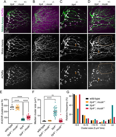
Musk depletion partially suppresses lrp4-mutant phenotype. (A,B) lrp4+/−;musk+/− trans heterozygotes have an innervation pattern indistinguishable from WT (A), whereas lrp4;musk double mutants phenocopy musk mutants with defasciculated axonal patterning labeled with Tg(mnx1:GFP) and diffuse AChR staining labeled by α-Btx (B). (C) lrp4-mutant motor axons have swellings in their innervation pattern that are opposed to large AChR clusters. (D) Although lrp4 mutants that are heterozygous for musk (lrp4−/−;musk+/−) still have some large AChR clusters similar to lrp4 mutants (orange arrows), they also have regions of the fin with smaller AChR clusters (encircled by orange line). (E-G) Quantification of the number of AChR clusters per fin (E), the median cluster size per fin (F) and the distribution of cluster sizes (5 μm2 bins) (G). ****P<0.001, one-way ANOVA with Sidak's (E) or Dunnett's (F) multiple comparisons test. n=8 (WT), 21 (lrp4+/−;musk+/−), 4 (lrp4−/−;musk−/−), 33 (lrp4−/−) and 39 (lrp4−/−;musk+/−). |

Agrin restricts presynaptic terminal and neural AChR cluster size. (A-E) Developmental time course from Tg(mnx1:GFP) larvae with motoneurons stained with α-Btx to label AChRs. (A,B) At 46 and 51 hpf, axons growing from the dorsal plexus (DP) have not yet innervated all prepatterned AChR clusters (single arrowheads). (C-E) Whereas small clusters are added as axons grow into the pectoral fin in sibling animals, clusters mainly increase in size in agrn mutants. Double arrow in D indicates presynaptic swelling colocalized with an AChR cluster. Only abductor innervation is shown, with the fin area outlined by the white-dashed line. Asterisks indicate endothelial or endoskeletal cells labeled in the green channel. (F) Schematic summarizing the developmental time course. Both siblings and agrn mutants look similar during the prepatterning stage. Incoming axons induce small AChR clusters in sibling animals, whereas, in agrn mutants, AChR clusters and axonal swellings increase in size over time. n=4-10 animals per genotype per time point. |