
- Title
-
Liposomal Clodronate-mediated Macrophage Depletion in the Zebrafish Model
- Authors
- Yang, L., Rojas, A.M., Shiau, C.E.
- Source
- Full text @ Bio Protoc

A. Capillary tube loaded into micro-needle puller. B. Magnified view of heated filament surrounding glass capillary. C. Creation of 2 microinjection needles from pulling (double-sided white arrow). |
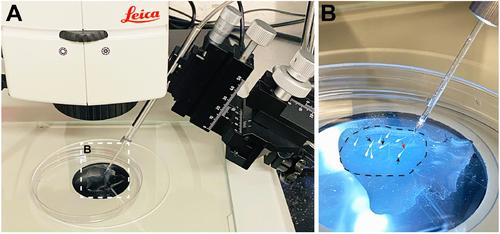
A. Mounted larvae are placed under a fluorescent stereomicroscope adjacent to a microinjection needle attached to a micro-manipulator. B. Higher magnification of mounted larvae. Dotted circle shows solidified thin layer of low-melt agarose covered with a small pool of system water supplemented with PTU. Black arrows point to larvae. Red arrow points to tip of needle (Note: This arrow is not pointing to the injection site.) |

A. Color of fish water after addition of neutral red. B. Color of fish and water after removal of neutral red. C. Full body image of control uninjected 4 dpf larvae after neutral red staining. D and E. Lateral (D) and dorsal (E) view of neutral red staining. C-E. Larvae imaged in 3% methyl cellulose. Black arrows point to individual microglial cells. |