- Title
-
Expression pattern of the small muscle protein, X-linked (smpx) gene during zebrafish embryonic and larval developmental stages
- Authors
- Ghilardi, A., Diana, A., Prosperi, L., Del Giacco, L.
- Source
- Full text @ Gene Expr. Patterns

ZFIN is incorporating published figure images and captions as part of an ongoing project. Figures from some publications have not yet been curated, or are not available for display because of copyright restrictions. EXPRESSION / LABELING:
|
|
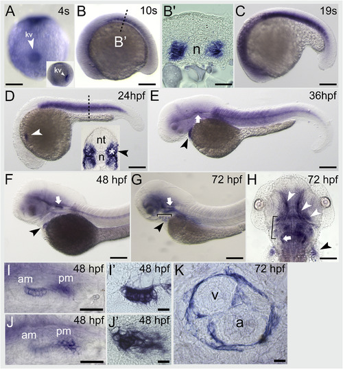
smpx embryonic expression pattern analyzed by in-situ hybridization. (A) At first smpx signal appears at 4-somite stage at the level of the Kupffer's vesicle (white arrowhead, frontal view), as also shown in the inset (lateral view). (B–D) From 10 to 24 hpf the gene is abundantly expressed in each somite. (B′) Cross-section of a 10 somites embryo showing smpx mRNA labeling the most adaxial cells next to the notochord. (D) The 24 hpf stage marks the onset of smpx expression in the developing heart (white arrowhead); smpx now labels both fast (white asterisk) and slow (black arrowhead) muscle fibers, as shown by the cross-section in the inset. (E–G) While smpx expression decreases in the trunk, the signal at 36, 48, and 72 hpf persists in the heart (black arrowhead), labeling both ventricle and atrium (K) and is now visible in the forming ear (white arrow), specifically in the territories corresponding to the anterior (I,I′) and posterior (J,J′) maculae. (G,H) Larvae at 72 hpf; besides heart and ear, smpx is also expressed in the muscles of the branchial arches (bracket), of the eye (white arrowheads) and of the pectoral fins (black arrowhead).B,C,D,E,F,G: Lateral views, anterior to the left. H: Dorsal view, anterior up. n, notochord; nt, neural tube; am, anterior macula; pm, posterior macula; v, ventricle; a, atrium. Scale bars = 200 μm in A,B; 20 μm in B’; 150 μm in C; 500 μm in D,E,F,G; 25 μm in I,J; 10 μm in I′,J′,K. EXPRESSION / LABELING:
|
|
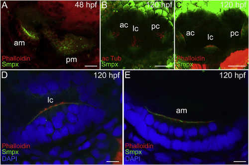
Smpx embryonic expression pattern analyzed by immunofluorescence. (A) Image of the anti-Smpx antibody staining the ear of a 48 hpf embryo and counterstained with phalloidin (F-actin) to visualize the cytoskeleton. Smpx signal is restricted to the region of the anterior and posterior maculae. (B,C) At 120 hpf Smpx is confined to the anterior, lateral and posterior cristae, labeling the apical membrane of the hair cells, below the kinocilia stained with the antibody against acetylated tubulin (B) and the stereocilia bundle painted with phalloidin (C). (D,E) Close-up views of the zebrafish ear at 120 hpf highlighting the lateral crista and the anterior macula; co-labeling of the apical membrane of the hair cells with the antibody against Smpx and with phalloidin; the nuclei (DAPI) are located in the basal portion of the cells. Images are all lateral views, anterior to the left, of confocal Z-stacks taken from whole mount embryos and larvae. am, anterior macula; pm, posterior macula; ac, anterior crista; lc, lateral crista; pc, posterior crista. Scale bars = 20 μm in A; 50 μm in B,C; 10 μm in D,E. |
|
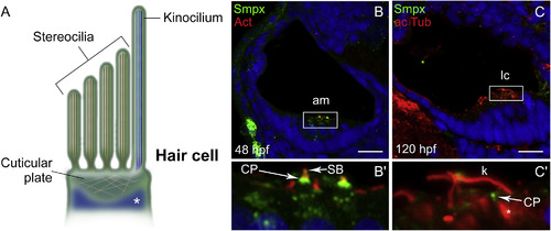
Smpx localizes to the cuticular plate of the inner ear hair cells. (A) Schematic representation of the hair cell apical membrane of the zebrafish inner ear; the kinocilium, with the structural tubulin organized in microtubules (in blue), and the bundle of F-actin-based mechanosensitive stereocilia (in brown) are depicted. The cuticular plate and the somatic tubulin (white asterisk) are also shown. (B,C) Paraffin sections co-labeled with antibodies against Smpx and phalloidin (B) and with antibodies against Smpx and acetylated tubulin (C), with the nuclei stained with DAPI. (B′,C′) 5X magnifications of B and C; Smpx is located in the region corresponding to the cuticular plate, in between the stereociliary F-actin-based bundle above (B′) and the somatic tubulin below (C′); F-actin (red) and Smpx (green) signals co-localize in the uppermost part of the cuticular plate (yellow in B′). Images are all lateral views, anterior to the left, of confocal Z-stacks taken from paraffin sections from embryos and larvae. am, anterior macula; lc, lateral crista; CP, cuticular plate; SB, stereociliary bundle; k, kinocilium. Scale bars = 30 μm. |
|
Smpx localizes to the ciliated cells of the zebrafish pronephros. (A) Cross-section of the trunk of a 48 hpf embryo; the pronephric ducts are labeled in green with the antibody against Smpx. (A′) 5X magnifications of the area enclosed in the rectangle in A; Smpx labels the apical membrane of the ciliated cells facing the lumen of the duct, as also confirmed (B) by the co-labeling with antibodies against Smpx and acetylated tubulin, where the lumen is populated exclusively by the acetylated tubulin-based kinocilia (red) sprouting from the apical membrane and devoid of Smpx (green). Images are all confocal Z-stacks taken from paraffin sections. pd, pronephric duct; n, notochord; nt, neural tube. Scale bars = 50 μm in A; 20 μm in C. EXPRESSION / LABELING:
|
Reprinted from Gene expression patterns : GEP, 36, Ghilardi, A., Diana, A., Prosperi, L., Del Giacco, L., Expression pattern of the small muscle protein, X-linked (smpx) gene during zebrafish embryonic and larval developmental stages, 119110, Copyright (2020) with permission from Elsevier. Full text @ Gene Expr. Patterns


