FIGURE SUMMARY
Title

CiliaCarta: An integrated and validated compendium of ciliary genes

Authors
van Dam, T.J.P., Kennedy, J., van der Lee, R., de Vrieze, E., Wunderlich, K.A., Rix, S., Dougherty, G.W., Lambacher, N.J., Li, C., Jensen, V.L., Leroux, M.R., Hjeij, R., Horn, N., Texier, Y., Wissinger, Y., van Reeuwijk, J., Wheway, G., Knapp, B., Scheel, J.F., Franco, B., Mans, D.A., van Wijk, E., Képès, F., Slaats, G.G., Toedt, G., Kremer, H., Omran, H., Szymanska, K., Koutroumpas, K., Ueffing, M., Nguyen, T.T., Letteboer, S.J.F., Oud, M.M., van Beersum, S.E.C., Schmidts, M., Beales, P.L., Lu, Q., Giles, R.H., Szklarczyk, R., Russell, R.B., Gibson, T.J., Johnson, C.A., Blacque, O.E., Wolfrum, U., Boldt, K., Roepman, R., Hernandez-Hernandez, V., Huynen, M.A.
Source
Full text @ PLoS One

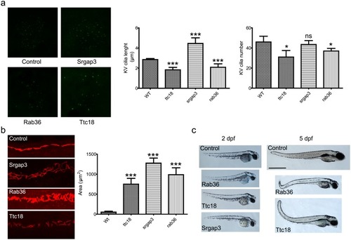
Validation by zebrafish phenotype.

A) Cilia length and number in zebrafish Kupffer’s vesicles. The length and number of cilia in ttc18 and rab36 morphants are significantly reduced (p<0.001 and p<0.05 resp.). Cilia in srgap3 morphants are elongated (p<0.001) but the number of cilia is normal. Bars represent mean ± S.E.M. B) Pronephric ducts in 24 hpf morphants. Cilia are stained with antibodies against acetylated alpha tubulin. The pronephric ducts are significantly enlarged for all three morphants compared to wild type (p<0.001). Bars represent mean ± S.E.M. C) Whole embryo phenotype 2 days post fertilization (dpf) and 5 dpf zebrafish control and morphant embryos. All morphants exhibit the body curvature that is characteristic for cilia dysfunction. Note that in our screening we did not manage to obtain surviving srgap3 morphants past 3 dpf.

OSCP1 localizes to the cilium and regulates ciliary function in vivo.

A) GFP-tagged OSCP-1 driven by its endogenous promoter is specifically expressed in ciliated sensory neurons in C. elegans, and the GFP-fusion protein is concentrated along the length of the cilium. Shown are fluorescent images of OSCP-1::GFP and XBX-1::tdTomato (ciliary marker) localization in amphid and phasmid cilia. Basal bodies (bb) and cilia are indicated. B) OSCP1::eCFP localization in hTERT-RPE1 cells. OSCP1 localizes to the basal body and daughter centriole as well as in the cytosol in a punctate manner. C) OSCP1 localization in human respiratory cells (red) co-stained with acetylated tubulin (green). OSCP1 localizes to the cytosol, but specifically to the base of the ciliated crown of these multi-ciliated cells. D) Indirect high magnification immunofluorescence of OSCP1 (green) and centrin-3 (Cen3, red), a marker protein for the connecting cilium (CC), the basal body (BB) and the adjacent centriole (Ce), in the photoreceptor cilium region of an adult mouse. Immunoelectron microscopy of CCDC113 confirms the localization of OSCP1 at the base of the cilium. The schematic represents a zoom of the ciliary region of a photoreceptor stained for OSCP1 and Cen3 in the according colors. IS, inner segment; OS, outer segment. E) Immunostaining of serum-starved murine IMCD3 cells with OSCP1 antibodies. OSCP1 is expressed in a punctate pattern along the axoneme. Acetylated tubulin and γ-tubulin (green), OSCP1 (Proteintech, 12598-1-AP, red) and nuclei (blue). Scale bar 5 μm. F) Zebrafish embryos injected with 4 ng of oscp1 splice morpholino 2 and 4 days post-fertilization (dpf). The characteristic ciliary phenotype with a curved body, small eyes and melanocyte migration defects at 2dpf. 4 dpf morphants display obvious pronephric cysts, small eyes, heart edemas, small heads and short bodies. Scale bar 500 μm. G) Left panel: details of the head of 2 dpf zebrafish embryos showing small eyes and small head in the oscp1 morphants. Scale bar 500 μm. Right panel: dorsal view of 4 dpf zebrafish embryos. Left embryo is a 4dpf oscp1 morphant. Right embryo is a control. Scale bar 200 μm. Note the small eyes, melanocyte migration defects and small fin buds (white arrows) in the oscp1 morphants compared with the control fin buds (black arrows). H) Immunofluorescence staining of pronephric cilia at 24 hpf (acetylated α-tubulin). 4 ng oscp1 morphants display shortened and disorganized cilia in the medial portion of the pronephric ducts. I) Detail of a pronephric cyst (outlined) in a 4 dpf zebrafish morphant. Scale bar 500 μm. J) Oscp1 morphants Kupffer’s vesicle cilia staining. Oscp1 morphants show smaller Kupffer’s vesicles with reduced cilia number per Kupffer’s vesicle (56 controls vs. 33 oscp1 morphants) and shorter cilia (controls; 7.1 μm vs oscp1 morphants; 5.7 μm. Significance was determined by t-test p-value<0.01. K) Dose dependent phenotype of oscp1 morphants. After injecting 4 ng of oscp1 morpholino the percentage of embryos with a weak phenotype is 51% and embryos with a strong phenotype is 28%. Those percentages change when 6 ng of morpholino are used with 31% of embryos showing with a weak phenotype and 27% with a strong phenotype, (strong and weak phenotypes described in S7 Fig). The number of dead embryos increases when 6ng of oscp1 morpholino are used (38%) compared with 4 ng of oscp1 morpholino (10%). We injected zebrafish embryos at one cell stage with 4 ng of oscp1 morpholino and 100 pg of human OSCP1 mRNA. The rescue increased the normal phenotype percentage from 8% to 43% and decreased the weak phenotype from 51% to 27% and the strong phenotype from 28% to 9.5%. Significance was determined by χ2 test, p<0.0001.

oscp1 zebrafish morpholino phenotypes.

Zebrafish strong oscp1 mo phenotype: short body, small eye, obvious pronephric cyst development, and obvious heart edemas. Zebrafish weak oscp1 mo phenotype: short body, small eye, small heads, but no obvious pronephric cyst or heart edemas.

Acknowledgments
This image is the copyrighted work of the attributed author or publisher, and ZFIN has permission only to display this image to its users. Additional permissions should be obtained from the applicable author or publisher of the image. Full text @ PLoS One