- Title
-
Central role of autophagic UVRAG in melanogenesis and the suntan response
- Authors
- Yang, Y., Jang, G.B., Yang, X., Wang, Q., He, S., Li, S., Quach, C., Zhao, S., Li, F., Yuan, Z., Lee, H.R., Zhong, H., Liang, C.
- Source
- Full text @ Proc. Natl. Acad. Sci. USA
|
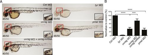
UVRAG is required for melanocyte development in zebrafish in vivo. (A) Representative images of 48 h postfertilization (hpf) zebrafish embryos injected with control morpholino (MO) or MO targeting uvrag, pmel, or tyr. Rescue of uvrag MO-mediated depigmentation is shown by coinjection of human uvrag mRNA. Box highlights area of enlargement visualized in Insets. Numbers on each panel indicate the number of embryos showing the representative phenotype per total number of embryos examined. (B) Quantitative measurement shows significantly decreased pigmentation in uvrag MO and pmel MO group compared with the control group. Means were calculated from the data collected from three independent experiments (n = 50). ***P < 0.001; ****P < 0.0001. PHENOTYPE:
|
|
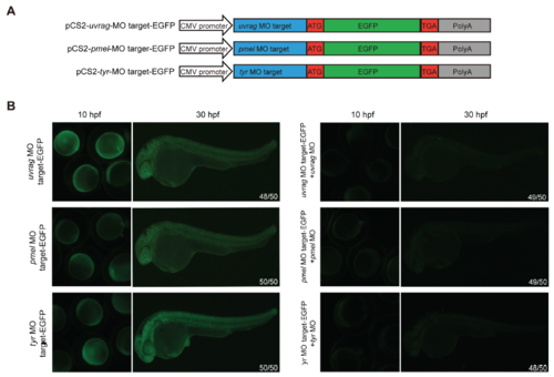
Related to Fig. 3: Effect of UVRAG deficiency on melanosome biogenesis in vivo. (A) Schematic representation of injected vectors containing the morpholino (MO) target sequence fused to GFP. The target site of uvrag, tyr, and pmel is fused to the 5’ of GFP. (B) Efficacy and specificity of uvrag, pmel,, and tyrosinase (tyr) MO. Left panels show Zebrafish embryos injected with the MO target-GFP fusion constructs as indicated. Right panels show co-injection of both MO target-GFP fusion constructs and MOs as indicated. Embryos were imaged at 10 hrs and 30 hrs post-fertilization (hpf) with the exposure time of 1.5 s in all groups. The disappearance of GFP expression (right panels) highlights the specific depletion of the target gene in Zebrafish embryos. |


