- Title
-
Shaping of Signal Transmission at the Photoreceptor Synapse by EAAT2 Glutamate Transporters
- Authors
- Niklaus, S., Cadetti, L., Vom Berg-Maurer, C.M., Lehnherr, A., Hotz, A.L., Forster, I.C., Gesemann, M., Neuhauss, S.C.F.
- Source
- Full text @ eNeuro
|
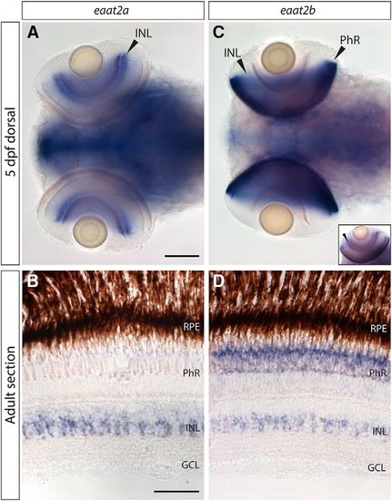
Transcript expression of excitatory amino acid transporter 2 (eaat2) paralogs. A, B, eaat2a mRNA is strongly expressed in the inner nuclear layer (INL) in the retina, in both 5-d postfertilization (5-dpf) larvae (A) and adult retina (B). Additionally, extremely low transcript levels can be found in photoreceptors (B). C, D, mRNA of eaat2b is expressed in photoreceptors and weakly in the INL throughout different developmental stages (C, in 5-dpf larvae; D, in adult retinal sections). Small inset in C shows eaat2b in situ staining in an eye of a whole-mount larva that has been only shortly stained, to better visualize expression in the INL. Scale bar in A is 100 µm; also applies to C. Scale bar in B corresponds to 50 µm; also applies to D. |
|
Protein expression of EAAT2 paralogs. A–F, Double immunostaining of EAAT2a (green) and glutamine synthetase (magenta) in adult (A) and larval (5 dpf; D) retinal sections confirm expression of EAAT2a in Müller glia cells. Separated channels are shown in B (adult), E (5 dpf; EAAT2a, green channel only), C (adult), and F (5 dpf; glutamine synthetase, magenta channel only). Scale bar in A is 30 µm; also applies to B and C. Scale bar in D is 50 µm; also applies to E and F. G–N, EAAT2b protein is expressed in a dotted manner in the outer plexiform layer (OPL) in all cone pedicles, but it is not expressed in rods. EAAT2b antibody staining (magenta) on adult retinal sections stained with Zpr-1 (red-green double cones, G) and on retinal sections of zebrafish expressing GFP in blue cones (H), UV cones (I), and rods (J) confirms that EAAT2b is cone specific and is spared from rod spherules. K–M show zoom-ins of the cone pedicles expressing EAAT2b (magenta) in red-green double cones (K), blue cones (L), and UV cones (M). N shows larval (5 dpf) expression of EAAT2b in magenta together with a nuclear counterstain (DAPI, blue). Scale bars in G–J are 7 µm. Scale bars in K–M are 2 µm. Scale bar in N is 30 µm. |
|
Confirmation of knockdown. A–C, Immunostaining of EAAT2a on WT (A) and EAAT2a morphant [B, 1.3 ng EAAT2a morpholino (MO) 1; C, 1.8 ng EAAT2a MO 2] retinal sections (5 dpf). D, Box-and-whisker plot of analysis of fluorescence of WT, EAAT2a MO 1, and EAAT2a MO 2 injected animals stained with anti-EAAT2a antibody. Statistical analysis reveals a highly significant (p < 0.001) reduction of fluorescence for both MOs. E–G, Retinal sections of WT (E) and EAAT2b morphant (F, 1.8 ng EAAT2b MO 1; G, 9 ng EAAT2b MO 2) larvae stained with anti-EAAT2b antibody. H, Fluorescence was measured in the OPL, and background fluorescence (taken from area in INL) was subtracted. Fluorescence of WT and morphant immunostaining is plotted in a box-and-whisker plot and shows a significant (p < 0.01) and slightly significant (p < 0.05) decrease in fluorescence in animals injected with 1.8 ng EAAT2b MO 1 and 9 ng EAAT2b MO 2, respectively. EAAT2a WT, n = 6; EAAT2a MO 1, n = 8; EAAT2a MO 2, n = 8; EAAT2b WT, n = 10; EAAT2b MO 1, n = 10; EAAT2b MO 2, n = 10. Scale bar in A is 30 μm; also applies to B and C. Scale bar in E is 10 μm; also applies to F and G. |
|
Retinal histology of EAAT2 morphant zebrafish larvae. Histologic analysis of retinal sections of WT, EAAT2a, and EAAT2b morphant zebrafish larvae (5 dpf) stained with Richardson–Romeis (A–C and G–I). Immunostaining of glutamine synthetase (green) labeling Müller glia cells counterstained with Bodipy (magenta; D–F) of WT and EAAT2a morphant (E, EAAT2a MO 1; F, EAAT2a MO 2) 5-dpf retinal sections. Anti–Zpr-1 immunostaining (labeling red and green cones, shown in green) on WT (J) and EAAT2b morphant (K, EAAT2b MO 1; L, EAAT2b MO 2) retinal sections counterstained with Bodipy (magenta). Knockdown of neither EAAT2a (B, EAAT2a MO 1; C, EAAT2a MO 2) nor EAAT2b (H, EAAT2b MO 1; I, EAAT2b MO 2) causes any defect in retinal lamination. Thickness of the retina was assessed on WT and morphant larvae and did not reveal any significant difference in the thickness of the retina in either EAAT2a or EAAT2b morphants (M, O), yielding p values of 0.997 and 0.935 for EAAT2a MO 1 and EAAT2a MO 2, respectively, and 0.658 and 0.922 for EAAT2b MO 1 and EAAT2b MO 2 (all in comparison to WT). Moreover, knockdown of EAAT2a does not significantly influence Müller glia cell length (N), nor does the loss of EAAT2b result in cone length alteration (P). Statistical analysis of the cell length yielded p values of 0.969 and 0.989 for EAAT2a MO 1 and EAAT2a MO 2, respectively, and 0.911 and 0.631 for EAAT2b MO 1 and EAAT2b MO 2 (in comparison to WT). All scale bars are 50 µm. Scale bar in A also applies to B and C; scale bar in D also applies to E and F; scale bar in G also applies to H and I; and scale bar in J also applies to K and L. |

ZFIN is incorporating published figure images and captions as part of an ongoing project. Figures from some publications have not yet been curated, or are not available for display because of copyright restrictions. PHENOTYPE:
|


