- Title
-
Polysaccharides from Ganoderma lucidum attenuate microglia-mediated neuroinflammation and modulate microglial phagocytosis and behavioural response
- Authors
- Cai, Q., Li, Y., Pei, G.
- Source
- Full text @ J Neuroinflammation
|
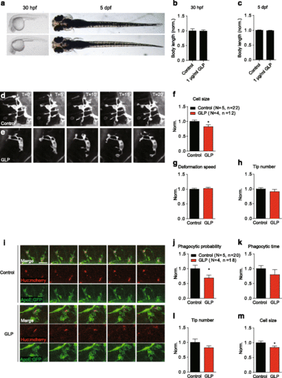
GLP modulates microglial morphology and phagocytosis in vivo. Zebrafish embryo (30 hpf) grown in GLP revealed no developmental defect (a). Continued treatment with GLP during the embryonic developmental stage did not affect the larva body lengths (b, c). Dorsal view of the optic tectum in a 5-dpf Tg(Apo-E:eGFP, HuC:mCherry) zebrafish larvae. Time-lapse images of the dynamics of resting microglia pre-treated with (e) or without (d) 1 μg/ml GLP. The time scales were in minutes. f–g The effects of GLP on resting microglial cell sizes, deformation speed and tip numbers. h Time-lapse images showing the phagocytic process of microglia Tg(Apo-E:eGFP, HuC:mCherry) zebrafish larvae at 5 dpf. The effect of 1 μg/ml GLP on activated microglial phagocytosis (j, k) and morphology (l, m). Data normalised to control. N stands for numbers of zebrafish and n for the number of cells. Statistical analysis was performed using unpaired two-tailed Student’s t test. Values expressed are mean ± SEM. (*p < 0.05) EXPRESSION / LABELING:
|

