- Title
-
Transcription factor Emx2 controls stereociliary bundle orientation of sensory hair cells
- Authors
- Jiang, T., Kindt, K., Wu, D.K.
- Source
- Full text @ Elife
|
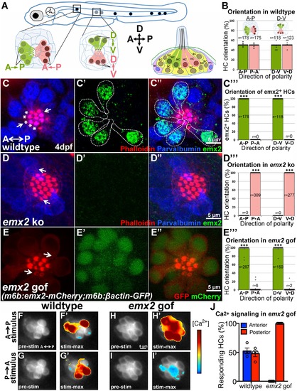
emx2 regulates hair bundle polarity in neuromasts. (A) Surface view of an A-P and D-V oriented neuromast and a sagittal view of the cellular architecture of a neuromast in the lateral line of zebrafish. (B) HCs with opposite orientation are in a 1:1 ratio. (C–C'') An example and (C''') quantification showing only HCs (parvalbumin-positive, blue) with hair bundles (phalloidin, red) oriented toward the posterior (C, asterisks) or ventral direction are positive for anti-Emx2 staining (green) in A-P and D-V neuromasts, respectively (55 neuromasts from 26 larvae, ***p<0.001; Figure 10—source data 1). (D–D'') An example and (D”') quantification of HCs in emx2 knockouts showing all HCs are emx2-negative (D') and pointing towards the same direction (36 neuromasts, 18 larvae, ***p<0.001; Figure 10—source data 1). (E–E''') All hair bundles (visualized using m6b:βactin-GFP, red) are Emx2-positive (E',E'', green) and pointing toward the posterior in the A-P oriented neuromasts of gof mutants. (E''') Quantification of hair bundle polarity in 43 A-P and D-V neuromasts from 23 larvae (Figure 10—source data 1). ***p<0.001. (F–J) In response to an anterior-to-posterior stimulus (F') or posterior-to-anterior stimulus (G') a similar percentage of wildtype HCs show mechanically-evoked increase in calcium levels (J; Figure 10—source data 2). Emx2 gof HCs only respond to an anterior-to-posterior stimulus (H') and are inhibited by a posterior-to-anterior stimulus (I'). The heatmap indicates the change in calcium levels during the first half of the 2s-step stimulus (2 larvae and 4 neuromasts in control, 2 larvae and 7 neuromasts in emx2 gof). Error bar represents SEM. EXPRESSION / LABELING:
PHENOTYPE:
|
|
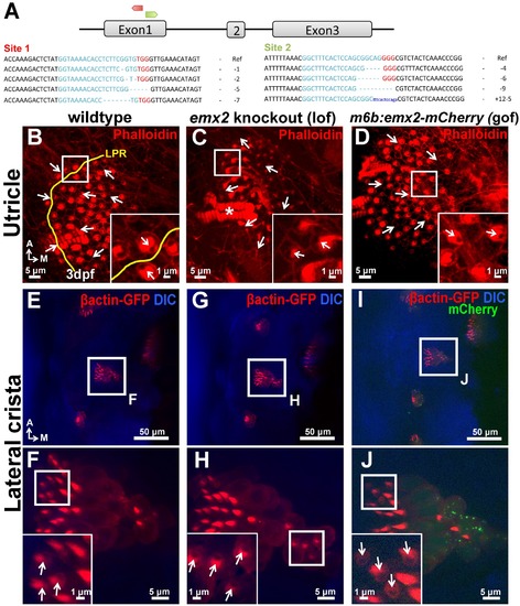
Polarity phenotypes in the utricle and lateral crista of emx2 loss- and gain-of function zebrafish mutants. (A) Two targeted sites of deletion (red site 1 and green site 2) in exon1 of emx2 using CRISPR/Cas9 technology. Guide targets (blue), PAM sequences (red), and examples of deletions found are shown. (B–D) Utricle and (E–J) lateral crista of controls (B,E,F), emx2 knockout (C,G,H), and emx2 gain-of-function, m6b:emx2-mCherry, zebrafish (D,I,J). (B–D) The LPR is by the lateral edge of the utricle in zebrafish (B; n = 10), which is absent in emx2 knockout utricles (C; n = 13) as well as in gof (m6b:emx2-mCherry) utricles (D; n = 9). Based on phalloidin staining (red), HCs at the edge of emx2 knockout utricles point toward the lateral (inset in C), whereas HCs in the medial region of gof utricles now point toward the medial (inset in D), opposite from controls (inset in B). (E–J) Arrows indicate the direction of hair bundle polarity based on βactin-GFP label (red). Hair bundles in the lateral crista of control are pointing toward the anterior direction (E,F; n = 6) and this polarity is not affected in the emx2 knockout fish (G,H; n = 3) but reversed in the gof cristae (I,J; n = 3). |


