- Title
-
Characterization of genetic loss-of-function of Fus in zebrafish
- Authors
- Lebedeva, S., de Jesus Domingues, A.M., Butter, F., Ketting, R.F.
- Source
- Full text @ RNA Biol.
|
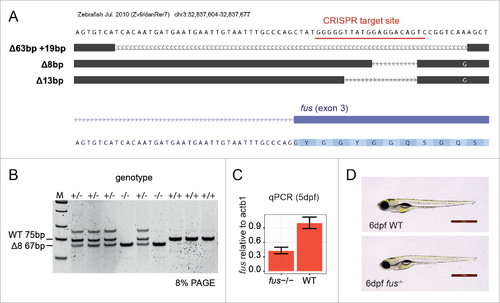
Generation and validation of the fus knockout zebrafish. (A) fus alleles generated by CRISPR-Cas9. A screenshot from the UCSC genome browser shows aligned sequences from heterozygous F1 animals. CRISPR target site in fus exon 3 is underlined in red. (B) An example of a PAGE gel for genotyping the Δ8 allele. M = Low molecular weight marker. Wild type (75bp) and fus−/− (67bp) products can be discriminated. (C) Quantification of fus mRNA relative to WT mRNA in 5dpf embryos. (D) Normal morphology of the WT and fus−/− embryo at 6 d post fertilization (dpf). Scale bar is 1mm. |
|
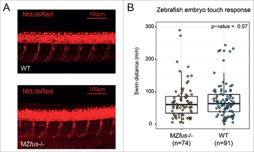
Motoneuron morphology and touch evoked escape response of fus knockout larvae. (A) Confocal images of trunks of 36hpf larvae expressing neuron-specific DsRed.28 Maternal zygotic Fus knockout larvae show normal overall primary motoneuron axon morphology indistinguishable from WT. Maximal intensity projection of a confocal stack is shown; scale bar is 100µm. (B) Touch evoked escape response of 2dpf (48–52hpf) larvae. Swim distances in mm for individual larvae tracks are plotted. P-value is from Kruskal-Wallis test. n indicates the number of larvae tested for each group. |
|
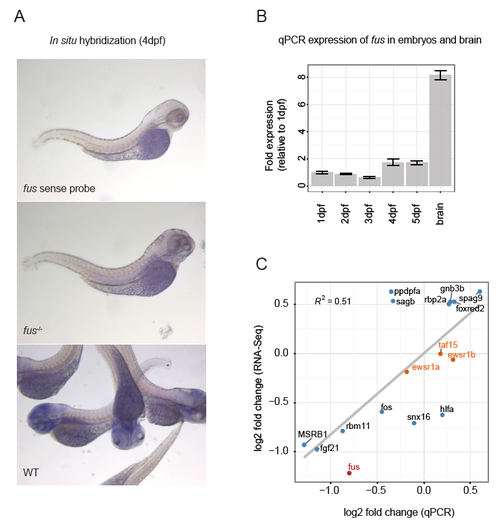
(A) In situ hybridization showing mRNA expression of fus mRNA in 4dpf larvae. (B) fus mRNA levels at several larval stages and in the adult brain of WT animals (qPCR) (C) qPCR validation of selected genes showing mRNA fold changes in RNA-Seq in the fus-/- vs WT. fus is downregulated (red), whereas other FET family genes are unchanged (taf15, ewsr1a, ewsr1b, orange) |
|
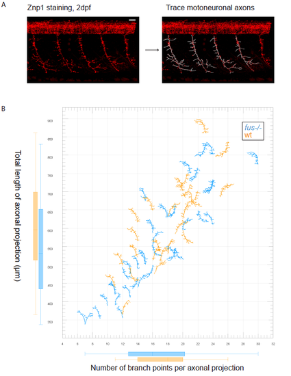
Analysis of axonal projection branching in wt and fus-/- 2dpf embryos with Imaris. (A) Example screenshots from the Imaris work area. The original trunk section is shown on the left, the same section with the overlay of traced motoneuronal projections to the right. Deconvolved images of Znp1 staining of trunk sections containing 4-5 axonal projections were used for analysis. Only one side of the trunk, either left or right, was imaged for clarity. Filaments package of Imaris was used for semiautomatic tracing of axonal projections. Scale bar is 20μm. (B) Scatterplot representing measurements from seven wild type and eight fus-/- trunk sections. In total, 36 fus -/- and 25 wt axonal projections were measured. Total length of axonal projection in μm is plotted against number of branching points per axonal projection. The plot was created with Vantage package of Imaris. Each data point is a representation of the corresponding Filament object. Starting point of the filament corresponds to the actual data point. Some filaments are inverted because the corresponding trunk was imaged in inverted orientation. Wt projections are shown in orange, fus-/- in blue. PHENOTYPE:
|

ZFIN is incorporating published figure images and captions as part of an ongoing project. Figures from some publications have not yet been curated, or are not available for display because of copyright restrictions. |


