- Title
-
SPP2 Mutations Cause Autosomal Dominant Retinitis Pigmentosa
- Authors
- Liu, Y., Chen, X., Xu, Q., Gao, X., Tam, P.O., Zhao, K., Zhang, X., Chen, L.J., Jia, W., Zhao, Q., Vollrath, D., Pang, C.P., Zhao, C.
- Source
- Full text @ Sci. Rep.
|
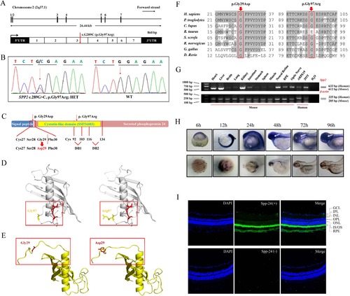
Genetic analyses of the SPP2 variants, and expression profiling of Spp-24. (A) SPP2 gene spanning 26.44 kb on chromosome 2q37.1 contains 7 exons (upper panel). The identified heterozygous variant, c.G289C (p.Gly97Arg), was located within exon 3 (below panel). (B) Sequencing chromatogram of patient AD02-III:3 shows the c.289G>C substitution. WT sequence is also shown. (C) Schematic representation of the relative linear location of the two SPP2 mutations in context of Spp-24 protein structure. The p.Gly97Arg is located in the cystatin-like domain, and the artificial mutation, p.Gly29Asp, is located at the last residue of signal peptide. The red dotted line denotes the cleavage site of the signal peptide. The two internal disulfide bonds are indicated by DB1 and DB2 respectively. (D–E) Structural modeling of Spp-24. The mutated residue 97 is located within the first disulfide bond loop of Spp-24 (D, cys92-103 disulfide bond is denoted by red color). The other substitution p.Gly29Asp would lead to the distortion of the signal peptide cleavage site in the mutant when compared with the WT monomer (E). (F) Conservation analyses of the mutated residues 29 and 97 of Spp-24 in multiple species, including human (H. sapiens), chimpanzees (P. troglodytes), dogs (C. lupus), cows (B. taurus), pigs (S. scrofa), rats (R. norvegicus), chickens (G. gallus), and zebrafish (D. rerio). Conserved residues are shaded. (G) Expression of SPP2 in multiple murine tissues and human cell lines, namely ARPE19 and HEK 293T (upper panel). Expression of β-actin serves as internal control (lower panel). (H) Zebrafish whole mount in situ hybridization reveals the ubiquitously expression of the spp2 transcript throughout the development of zebrafish. A relatively higher expression in the eye wall after 48 hours post fertilization is indicated by white arrows. (I) Immunostaining for spp-24 (green) on murine retinal frozen section. Abundant reactivity of spp-24 was detected in the inner/outer segments (IS/OS) and retinal pigment epithelium (RPE) layers, and moderate staining was also found in sclera, choroidal vessels and ganglion cells (GC) (upper panel). Sections incubated with secondary antibody alone serve as negative control (lower panel). Scale bar: 100 µm. |

