- Title
-
Technical brief: Constant intense light exposure to lesion and initiate regeneration in normally pigmented zebrafish
- Authors
- Rajaram, K., Summerbell, E.R., Patton, J.G.
- Source
|
Effects of constant intense light exposure on cone cells of pigmented AB zebrafish. Retinas were collected from zebrafish before (0 h) and after the indicated length of light exposure. Retinas were processed for immunohistochemistry and stained with zpr1 antibody (red). Nuclei were counter-stained with TOPRO (blue). Dorsal retinas are shown. The arrowheads at 0 h indicate the ordered arrangement of the double cones; the bracket indicates ONL. White arrows show condensed zpr+ cells at 72 h of intense light. ONL, outer nuclear layer; INL, inner nuclear layer; GCL, ganglion cell layer. Scale bar is 50 µm. |
|
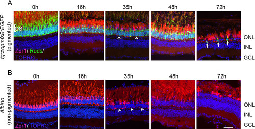
Photoreceptor loss in pigmented and nonpigmented retinas. Pigmented tg:zop:nfsB:EGFP zebrafish (A) or nonpigmented albino zebrafish (B) were subjected to intense light damage, and retinas were collected at the indicated time points and processed for immunohistochemistry. Retinas in (A) were stained with zpr-1 (red) and anti-green fluorescent protein (GFP; green) antibodies, while retinas in (B) were stained with zpr-1 (red) antibody; nuclei were counter-stained with TOPRO (blue). Dorsal retinas are shown. Arrowheads indicate regions of photoreceptor loss. Arrows denote rod photoreceptors that have rounded up and lost their outer segments. ONL, outer nuclear layer; INL, inner nuclear layer; GCL, ganglion cell layer; OS, outer segments. Scale bar is 50 µm. |
|
Terminal deoxynucleotidyl dUTP nick end labeling (TUNEL) labeling of light-treated pigmented retinas. Retinas from light-treated AB fish were collected at different times during light exposure and processed for TUNEL labeling (red). Nuclei were counterstained with TOPRO (blue). Scale bar is 50 µm. PHENOTYPE:
|
|
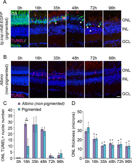
Time course of photoreceptor apoptosis in pigmented and nonpigmented retinas. Pigmented tg:zop:nfsB:EGFP zebrafish (A) or nonpigmented albino zebrafish (B) were subjected to intense light damage, and retinas were collected at the indicated time points and processed for terminal deoxynucleotidyl dUTP nick end labeling (TUNEL) labeling. A: Rods were stained with anti-green fluorescent protein (GFP; green) antibody; nuclei were counterstained with TOPRO (blue). Dorsal retinas are shown. Arrowheads indicate rods that appear condensed and lack OS. C: Quantification of TUNEL labeling for albino and pigmented zebrafish. Data represent mean±standard deviation (SD); *, p<0.005, n = 3-7 retinas for each time point. D: Quantification of the ONL thickness for albino and pigmented zebrafish. All comparisons were made at 0 h. Data represent mean±SD; *, p<0.05 using one-way analysis of variance with Tukey’s honest significant difference post hoc test; n = 5-7 retinas for each time point. ONL, outer nuclear layer; INL, inner nuclear layer; GCL, ganglion cell layer; OS, outer segments. Scale bar is 50 µm. |
|
Proliferating cell nuclear antigen (PCNA) expression during intense light exposure. Pigmented tg:zop:nfsB:EGFP zebrafish (A) or nonpigmented albino zebrafish (B) were subjected to intense light damage, and retinas were collected at the indicated time points and processed for immunohistochemistry. Retina sections were stained with anti-green fluorescent protein (GFP; green in panel A) and anti-PCNA (panels A and B) antibodies; nuclei were counterstained with TOPRO (blue). Dorsal retinas are shown. Arrowheads indicate PCNA+ rod precursors in the ONL. C: Quantification of PCNA+ cells in the INL and ONL. Data represent mean±standard deviation; n = 3-7 retinas per time point. ONL, outer nuclear layer; INL, inner nuclear layer; GCL, ganglion cell layer. Scale bar is 50 µm. EXPRESSION / LABELING:
|
|
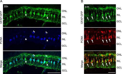
Light treatment of pigmented retinas stimulates proliferation of MG and progenitor cells. tg:GFAP:GFP fish were exposed to intense light for 35 h (A) or 51 h (B), and retinas were processed for immunohistochemistry. A: Proliferating cell nuclear antigen (PCNA) immunohistochemistry (blue) reveals co-staining with GFP+ MG in the central-dorsal retina. Few MG do not express PCNA (arrowheads in A). PCNA also labels rod precursors in the ONL (double arrowheads). B: Most GFP+ MG continue to express PCNA. Arrows indicate MG associated with small PCNA+ progenitor clusters. Few PCNA+ cells are migrating to the ONL. ONL, outer nuclear layer; INL, inner nuclear layer; GCL, ganglion cell layer. Scale bars are 50 µm. EXPRESSION / LABELING:
|
|
Light damage triggers tuba1a:GFP transgene expression in Müller glia (MG). A: tg:1016tuba1a:GFP zebrafish were exposed to the constant intense light paradigm, and retinas were collected at the indicated time points and assessed for GFP expression (red) by immunohistochemistry. GFP+ cells began to appear in the INL by 24 h (arrowheads). By 72 h, clusters of GFP+ cells were detected in the INL and ONL (double arrowheads). B: Immunohistochemistry for GFP and GS in 51-h light-exposed 1016tuba1a:GFP retinas. Arrows indicate GS+ and GFP+ MG. ONL, outer nuclear layer; INL, inner nuclear layer; GCL, ganglion cell layer; IPL, inner plexiform layer. Scale bars are 50 µm. EXPRESSION / LABELING:
|
|
Proliferating progenitor cells express tuba1a:GFP transgene following light damage. tg:1016tuba1a:GFP zebrafish were exposed to the constant intense light paradigm, and retinas were collected at the indicated time points and assessed for green fluorescent protein (GFP, green) and proliferating cell nuclear antigen (PCNA, red) expression in the central-dorsal retina by immunohistochemistry. Small clusters of GFP+/PCNA+ cells associate with MG processes at 51 h (double arrowheads). Large clusters of GFP+/PCNA+ cells span in the INL at 72 h (arrows). Arrowheads indicate rod precursors in the ONL. ONL, outer nuclear layer; INL, inner nuclear layer; GCL, ganglion cell layer; IPL, inner plexiform layer. Scale bar is 50 µm. EXPRESSION / LABELING:
|


