- Title
-
Negative feedback regulation of Wnt signaling via N-linked fucosylation in zebrafish
- Authors
- Feng, L., Jiang, H., Wu, P., Marlow, F.L.
- Source
- Full text @ Dev. Biol.
|
slc35c1 enhances the level of N-linked fucosylation expression in zebrafish embryos. (A) Schematic illustration of fucose biosynthetic pathways. Slc35c1 transports GDP-Fuc into the late secretary pathway (mainly the Golgi apparatus). (B) slc35c1 mRNA expression during zebrafish development as quantified by qPCR. The blue bars show slc35c1 relative expression during development and the red dots show the qPCR cycle number of the control gene odc1. (C) Western blot analysis of lysates of 5hpf zebrafish embryos expressing mouse Slc35c1-myc fusion protein. (D) Western blot analysis of global fucosylation levels at shield stage (6hpf) in embryos microinjected with gmds MO or mouse slc35c1-myc mRNA in the presence or absence of GDP-Fuc before and after PNGase F digestion. Proteins were resolved by SDS-PAGE and IB with AAL followed by anti-biotin-HRP in (D) or by streptavidin-Alexa488 for the quantification in (E). N-fucosylation: N-linked fucosylation; O-fucosylation: fucose in mucin and O-fucosylated proteins. EXPRESSION / LABELING:
PHENOTYPE:
|
|
Elevated N-fucosylation dorsalizes zebrafish embryos. (A) Variable degrees of dorsalization in embryos over-expressing mSlc35c1. (B) Quantification of the severity and penetrance of dorsalization in each treatment. (C and D) qPCR of the ventral transcriptional factors vox and vent in WT, GDP-Fuc injected or mSlc35c1 expressing embryos. (E–L) in situ hybridization with dorsal (chordin and gsc) and ventral (eve1) specific markers at shield stage (6hpf). (E–H) chordin expression in treated embryos (I–L) eve1 and gsc expression. The expression angles of chordin and gsc of embryos with each treatment are summarized in (M) and (N) respectably. The sample sizes for each treatment are as follows: wt (N=15), GDP-Fuc(N=13), mSlc35c1(N=18) and mSlc35c1 plus GDP-Fuc (N=21) for (M); and wt (N=19), GDP-Fuc (N=15), mSlc35c1(N=21) and mSlc35c1 plus GDP-Fuc (N=22) for (N). For chordin angle, ordinary One-way ANOVA between sample group means, p=0.0002. For gsc angle, ordinary One-way ANOVA between sample group means, p=0.0001. Note: ** Tukey′s procedure after ANOVA p<0.01. Null hypothesis for Χ2 between embryos injected with 1 ng mSlc35c1 and with GDP-Fuc plus 1 ng mSlc35c1 p=1.41E-04; for Χ2 between embryos injected with 2 ng mSlc35c1 and with GDP-Fuc plus 2 ng mSlc35c1 p=8.99E-03. EXPRESSION / LABELING:
|
|
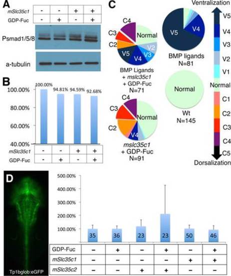
BMP and Notch signaling are intact in mSlc35c1 expressing embryos. (A) Phospho-Smad1/5/8 antibody blot indicates BMP signaling activity in each treatment. (B) Quantification of Phospho-Smad1/5/8 levels, normalized to α-Tubulin protein levels. (C) Molecular interaction between BMP signaling and Slc35c1 function. (D) Dorsal view of a transgenic zebrafish larva expressing a GFP reporter of Notch activity. Graph shows the GFP expression levels determined by the fluorescence intensities of transgenic notch reporter embryos injected with the specified molecules. The data represent the average of GFP reporter intensity from individual treated embryos, with WT set as 100%. |
|
GDP-Fuc or mSlc35c1 overexpression inhibits maternal Wnt signaling. (A-D) in situ hybridization of a direct Wnt signal target gene bozozok(dharma) in sphere stage (4hpf) embryos; lateral views. (E) Quantification of bozozok domain angle (e.g. dashed lines in A-D) in each group. The sample sizes for each treatment were as follows: gmds MO (N=35), wt (N=18), GDP-Fuc(N=54) and mSlc35c1(N=26). Ordinary One-way ANOVA between sample group means, p<0.0001. (F) qPCR of bozozok, a direct Wnt target gene in WT, GDP-Fuc injected or mSlc35c1 expressing embryos. bozozok expression is normalized to expression of the eef1a1a house keeping gene. (G) Schematic illustration of an embryo demonstrates the boxed regions shown in (H-K) for β-catenin protein localization at 512 cell stage (3hpf). View of the dorsal margin, as indicated by the nuclei of the yolk syncitial layer, which lack membranes (red arrowhead) at the margin, and the strong nuclear label (white arrowhead) in cells within the “core” region (i.e. closest to the Wnt8a source). The nuclear localization of β-catenin in the core region of (H) WT (n=6) and (I) mSlc35c2 (n=5) injected embryo, but is faint in either (J) GDP-Fuc (n=7) or (K) mSlc35c1 (n=9) injected embryos. Note: ** Tukeyós procedure after ANOVA **p<0.01; * p<0.05. Bar=100 μm. EXPRESSION / LABELING:
|
|
mSlc35c1 and GDP-Fuc can suppress Wnt8a induced patterning defects. (A–D) in situ hybridization of bozozok expression in injected embryos at sphere stage (4hpf). (E) Quantification of bozozok angle (area outlined by dashed lines in A–D). (F) qPCR of bozozok, a direct Wnt target gene, in embryos injected with wnt8a alone or combined with GDP-Fuc, gmds MO injected or mSlc35c1 expressing embryos. bozozok expression was normalized to the expression of the house keeping gene eef1a1a. (G) Posteriorization caused by over-expression of wnt8a. All embryos shown are 18hpf. P1-P4 denote the degree of posteriorization, as determined by the marker gene pax2a-krox20-myoD expression patterns. The P1 class lacks the telencephalon as indicated by loss of anterior pax2a expression, (red arrow). P2 embryos lack regions anterior to the MHB domain of pax2a, (red arrowhead). P3 embryos lack MHB expression of pax2a. In the P4 class krox20 expression indicates loss of anterior hindbrain segments. (H) Quantification of each treatment. Note: ** Tukey′s procedure after ANOVA p<0.01. Null hypothesis for Χ2 between embryos injected with wnt8a and with wnt8a plus mSlc35c1 p=6.05E-08. |
|
Excess fucosylation changes the response pattern of Wnt signaling. (A-B) Projections of stacks of individual confocal image sections show the nuclear localization of β-catenin in early zebrafish embryos (512 cell stage). (a–f) Single confocal sections. (A) β-catenin in a representative wild-type embryo. (a) Peripheral blastomeres at the animal pole. (b) medial region of the blastoderm. (c) The “core” region of highest Wnt activity at the dorsal margin. (B) The Wnt gradient of a representative mSlc35c1 expressing embryo. (d) The edge, (e) the medial and (f) the core and margin regions. (C-D) Schematic illustration shows Wnt activities based on β-catenin nuclear localization. In wild-type embryos, with the normal level of N-fucosylation, Wnt protein creates a gradient. In Slc35c1 over-expressing embryos, with excess N-fucosylation, Wnt protein levels are diminished, which reduces the highest Wnt activity response region; Lrp6 protein level is increased, which increases the basal level of the Wnt response, creating an extended zone of intermediate Wnt response. Note: Bar=200 µm. EXPRESSION / LABELING:
|
|
The interplay of Wnt8a signaling, slc35c1-mediated GDP-Fuc transport and N-linked fucosylation. (A) qPCR results show the relative expression of slc35c1 and slc35c2 in zebrafish embryos treated with wnt8a or DN-wnt8a mRNA and untreated control sphere stage (4hpf) embryos. All results are normalized to odc1. Expression in WT is defined as 100%. (B-C) Confocal images of slc35c1 fluorescent in situ show positive correlation between slc35c1 expression and β-catenin. Red: β-catenin protein staining. Green: in situ hybridization using slc35C1 antisense probe in (B) or slc35c1 sense probe in (C). Blue: nuclei. (D) A schematic model depicts the relationship between Slc35c1 and Wnt8a. Wnt8a activated canonical Wnt signaling enhances slc35c1 expression. Increased slc35c1 enhances N-linked fucosylation, which inhibits Wnt/β-catenin/TCF signaling during both blastula and gastrula stages. Therefore, these components comprise a negative regulatory loop to control Wnt signaling through N-fucosylation. Note: Bar=300 µm. EXPRESSION / LABELING:
|
Reprinted from Developmental Biology, 395(2), Feng, L., Jiang, H., Wu, P., Marlow, F.L., Negative feedback regulation of Wnt signaling via N-linked fucosylation in zebrafish, 268-86, Copyright (2014) with permission from Elsevier. Full text @ Dev. Biol.


