- Title
-
The Hippo Pathway Controls a Switch between Retinal Progenitor Cell Proliferation and Photoreceptor Cell Differentiation in Zebrafish
- Authors
- Asaoka, Y., Hata, S., Namae, M., Furutani-Seiki, M., Nishina, H.
- Source
- Full text @ PLoS One
|
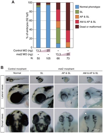
Mst2 is essential for early zebrafish embryogenesis. (A) Early developmental abnormalities of mst2 morphants. Control or mst2 morpholino (MO) at the indicated dose was injected into zebrafish embryos and phenotypes were analyzed at 52 hpf. Embryos were classified into five color categories on the basis of their phenotypes: blue, normal embryos; green, short body length (SL); orange, abnormal eye pigmentation (AP) accompanied by SL; red, abnormal eye morphology (AM) plus AP plus SL; and brown, dead or malformed embryos. Results are presented as the percentage of the total number of embryos examined (N). (B) Representative control and mst2 morphants at 52 hpf. Embryos were injected with control MO (13.3 ng) or mst2 MO (13.3 ng). Top panels, lateral views of whole embryos. Middle panels, higher magnification images of the head regions of the embryos in the top panels. Bottom panels, dorsal views of the head regions of the embryos in the top panels. (The head is at the top of each panel.) White arrowhead, representative area of AM. PHENOTYPE:
|
|
Forced expression of mRNA encoding constitutively active yap alters early zebrafish embryogenesis. (A) Representative images of EGFP mRNA-injected (control) or yap (WT) mRNA-injected zebrafish embryos at 52–54 hpf. Top panels, lateral views of whole embryos. Bottom panels, higher magnification images of the head regions of the embryos in the top panels. N, total number of embryos examined. Embryos injected with either yap (WT) mRNA or EGFP mRNA had normal phenotypes. (B) Representative images of EGFP mRNA-injected (control) or yap (5SA) mRNA-injected zebrafish embryos at 48 hpf. Embryos injected with Yap (5SA) mRNA (10 pg) showed the same spectrum of abnormal phenotypes as mst2 morphants. Data are presented as for Fig. 1B. |
|
Yap is directly involved in zebrafish retinogenesis. (A) Schematic illustration of the base hsp70-EGFP-yap construct (left panel) and the procedure for the heat shock experiment (right panel). Zebrafish embryos at the one-cell stage were injected with plasmid DNA containing the heat shock promoter constructs indicated in (B). At 21 hpf, injected embryos were immersed in a 37°C water bath for 1 h to apply heat shock and thus induce expression of EGFP-fused Yap. At 54 hpf, EGFP-expressing embryos were isolated and classified on the basis of their phenotypic features. (B) Representative images of the embryos in (A) that were injected with heat shock promoter constructs as indicated on the left side of panels. For each column, top right panels show lateral views of whole embryos, top left panels show higher magnification images of the head regions of the embryos, and bottom panels are fluorescent images of the corresponding top panels. White arrowheads, areas of AP. (C) Quantification of phenotypes of the embryos injected with heat shock promoter constructs in (A, B) as analyzed at 54 hpf. Color classification is as for Fig. 1A except that the phenotype of AP alone is indicated by striped orange shading. Results are presented as the percentage of the total number of embryos examined (N). |
|
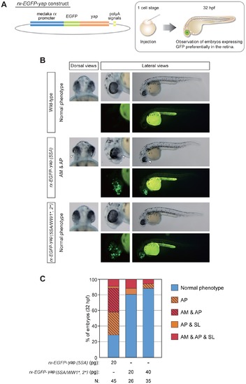
Retina-specific expression of yap (5SA) induces retinogenesis defects without affecting body axis formation. (A) Schematic illustration of the base rx-EGFP-yap construct (left panel) and the procedure for the experiment (right panel). Zebrafish embryos at the one-cell stage were injected with plasmid DNA containing the rx promoter constructs indicated in (B). (B) Representative dorsal and lateral views of the embryos in (A) that were injected with rx promoter constructs as indicated on the left side of panels. Data are presented as for Fig. 4B. White arrowheads, areas of AM plus AP. (C) Quantification of phenotypes of the embryos injected with rx promoter constructs in (A, B) as analyzed at 32 hpf. Color classification is as for Fig. 4C except that the phenotype of AM plus AP is indicated by striped red shading. Results are presented as the percentage of the total number of embryos examined (N). Note that expression of yap (5SA) variants mutated in both the WW1 and WW2 domains prevented the appearance of abnormal eye phenotypes. |
|
Morphological analysis of yap (5SA) mRNA-injected zebrafish embryos during the gastrulation and segmentation periods. (A) Alignment of amino acid sequence of medaka Yap with its zebrafish homolog performed as in Fig. S1. *, conserved serine residues phosphorylated by Lats. (B) Representative images of yap (5SA) mRNA-injected zebrafish embryos (N = 3) at the indicated developmental stages during gastrulation. Embryos were injected with EGFP mRNA as a control. (C) Representative lateral images of the embryos in (B) examined at the indicated stages during segmentation. |


