- Title
-
Isolation and Genetic Characterization of Mother-of-Snow-White, a Maternal Effect Allele Affecting Laterality and Lateralized Behaviors in Zebrafish
- Authors
- Domenichini, A., Dadda, M., Facchin, L., Bisazza, A., and Argenton, F.
- Source
- Full text @ PLoS One
|
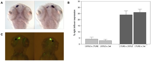
Zebrafish brain asymmetries are reversed in embryos from TLRE line. A, in situ hybridization on 3 dpf embryos showing the expression of the leftover (lov) gene, a marker of habenular L-R asymmetries. Normal lov expression is stronger in the left habenular nucleus, while in larvae with reversed asymmetries lov expression is stronger in the right habenula. B, frequencies of embryos with reversed (right) lov expression in embryos derived from GTLE females mated TLRE or wt males males and from TLRE females mated to GTLE or wt males. C, in vivo detection of the position of the parapineal organ (asterisk) in transgenic tg(foxD3:GFP)zf15 zebrafish. GFP expressed in the pineal complex allows discriminating between left parapineal (LEFT PPO) and right parapineal (RIGHT PPO) fish. EXPRESSION / LABELING:
|
|
Msw allele randomize Nodal pathway. A, From left to right, dorsal view of left-sided, bilateral and right-sided expression of lefty1 in the dorsal diencephalon detected in 22 somite-stage embryos. B, Percentages of normal (left-sided), bilateral and reversed lefty1 in WT control (n = 44), embryos from msw+/+ females (n = 33), embryos from msw+/- females (n = 435) and from msw-/- females (n = 403). C, spaw expression in the dorsal diencephalon in 15–18 somite-stage embryos. From left to right, dorsal view showing normal (left-sided), bilateral and right-sided expression. D, Percentages of normal (left-sided), bilateral and reversed spaw in WT control (n = 169), msw+/+ females (n = 126), embryos from msw+/- females (n = 203) and from msw-/- females (n = 753). PHENOTYPE:
|
|
Msw allele influence KV morphogenesis. A, zebrafish wt embryo at the 10-somite stage. Kupffer′s vesicle facing upwards (white arrowhead) is visible at the tail bud at the end of the notochord (n). The other panels show normal, reduced, and no Kupffer′s vesicle in embryos at the 10-somite stage. Scale bar = 50 μm. B, measures of area of KV in wt control embryos and in embryos derived from females of the three analyzed classes expressed as box plot (whiskers represent smaller and larger values for each group). Mean±SE are expressed. C, significative reverse correlation between the size of KV of embryos derived from two msw+/+, three msw+/-, and two msw-/- females and the frequency of larvae with reversed brain asymmetries generated by the same females. PHENOTYPE:
|
|
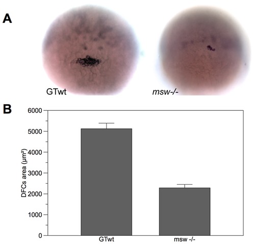
Msw allele seems to affect DFCs differentiation. A, sox17 expression at 50% epiboly in wt (left) and msw-/- embryo (right). B, measure of the area of DFCs cell mass at the 50% epiboly-stage in wt and msw-/- embryos. Mean±SE are expressed. PHENOTYPE:
|


