- Title
-
Zebrafish RNase T2 genes and the evolution of secretory ribonucleases in animals
- Authors
- Hillwig, M.S., Rizhsky, L., Wang, Y., Umanskaya, A., Essner, J.J., and Macintosh, G.C.
- Source
- Full text @ BMC Evol. Biol.
|
Characterization of zebrafish RNases. A) Ribonuclease activities present in zebrafish extracts. Adult zebrafish extracts were analyzed in an in gel RNase assay at two different pHs. Adult zebrafish of mixed sexes were separated into "body" (B, mostly muscle, skin and skeleton), "head" (H, which included skull, muscle, skin, brain, eyes among other tissues) and "gut" (G, which included most internal organs such as gut, liver, sexual organs, heart). The size range for RNase T2 and RNase A proteins is indicated. B) Same samples as in A, analyzed by SDS-PAGE and stained with Coomassie Blue. One hundred μg of protein per lane were analyzed in both types of gels. |
|
Expression of zebrafish RNase Dre1 and RNase Dre2. A) RT-PCR analysis of expression of RNase Dre2 and RNase Dre1 in adult tissues: B, brain; E, eye; H, heart; L, liver; G, gut; M, muscle; O, ovary; T, testis; S, skin. p70 was used as control for loading. B) RT-PCR analysis of expression of RNase Dre2 and RNase Dre1 in embryos at different times (in days) after fertilization. C) Ribonuclease activities present in zebrafish embryos (E) and adults (A) analyzed by in gel activity assay as in Figure 1. |
|
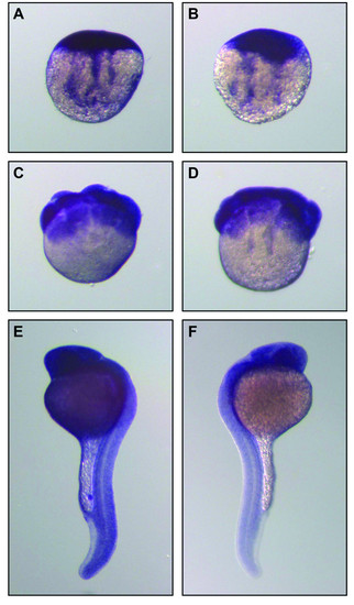
Localization of RNase Dre1 and RNase Dre2 expression in zebrafish embryos. Whole-mount in situ hybridization analysis was performed in embryos at the 1-cell stage (A, B), 16-cell stage (C, D) and prim 6 stage (E, F). Left panels, RNase Dre2 probe; right panels, RNase Dre1 probe. |


