- Title
-
Double-stranded RNA-activated protein kinase PKR of fishes and amphibians: Varying the number of double-stranded RNA binding domains and lineage-specific duplications
- Authors
- Rothenburg, S., Deigendesch, N., Dey, M., Dever, T.E., and Tazi, L.
- Source
- Full text @ BMC Biol.
|
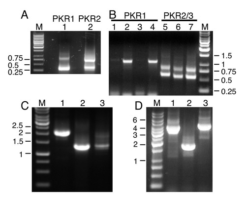
PCR for cloning of T. nigroviridis PKR genes. (A) Results for 5′ RACE PCRs with T. nigroviridis cDNA are shown for primers specific for PKR1 (lane 1) and PKR2 (lane 2). M denotes the 1 kb marker. Fragments are labeled in kb. (B) shows the results of 3′ RACE PCR using four different forward primers used in primary and nested PCRs for PKR1 (lanes 1–4) and three different forward primers combinations for PKR2 and PKR3 (lanes 5–7). The smaller fragment represents PKR2 and the larger one represents PKR3. (C) PCR products are shown using primers covering the complete open reading frames of PKR1 (lane 1), PKR2 (lane 2) and PKR3 (lane 3). (D) PCR reactions of overlapping regions were performed with genomic DNA to elucidate the genomic organization of PKR1. Lanes 1 and 2 show the PCR products obtained with primers spanning the region between the 5′ untranslated region of PKR1 and exon 15 and exon 14 and the 3′ untranslated region of exon 19, respectively. PCR product shown in lane 3 was obtained with primers covering exon 17 of PKR1 and exon 7 of PKR2. PCR products were cloned and completely sequenced. |
|
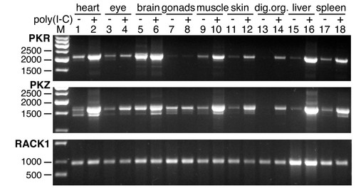
Comparison of expression patterns of zebrafish PKR and PKZ after induction with poly(I:C). PCRs were performed with cDNA prepared from the indicated tissues with primers covering the complete ORFs of zebrafish PKR (upper panel), PKZ (middle panel) or RACK1, the latter of which is constitutively expressed and served as control (lower panel). Zebrafish were either treated with poly(I:C) (indicated by plus) or with PBS (minus). EXPRESSION / LABELING:
|
|
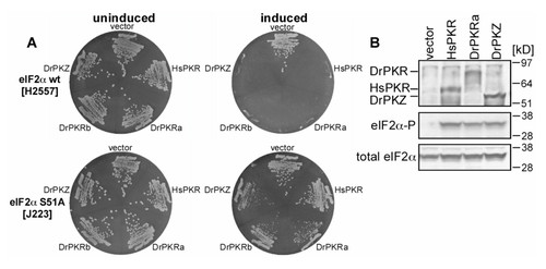
Both zebrafish PKR and PKZ phosphorylate yeast eIF2α. (A) Plasmids expressing full-length human (hs) PKR or zebrafish (dr) PKZ or two different alleles of PKR, under the control of a galactose-inducible promoter, were introduced into S. cerevisiae strains H2557 (wild-type eIF2α, upper plates) and J223 (eIF2α-S51A, lower plates) as indicated. After two rounds of single colony purification, the transformants were streaked out simultaneously on SD-ura (non-inducing conditions, left) or SGal-ura (inducing conditions, right) medium and grown for three days (SD-ura) or four days (SGal-ura). (B) Western blot analyses of extracts from strain H2557 transformed with vector, HsPKR, DrPKR or DrPKZ. SDS-PAGE was used to separate 4 μg of WCEs which were blotted onto nitrocellulose membranes. Tagged PKR and PKZ were detected using anti-Flag-tag antibody (upper panel). eIF2α phosphorylated on Ser51 and total eIF2α are shown in middle and bottom panels, respectively. |


