
- Title
-
Nonapical symmetric divisions underlie horizontal cell layer formation in the developing retina in vivo
- Authors
- Godinho, L., Williams, P.R., Claassen, Y., Provost, E., Leach, S.D., Kamermans, M., and Wong, R.O.
- Source
- Full text @ Neuron
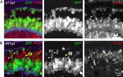
HC Precursors Express Progenitor Markers (A) Immunostaining of a retinal cross-section from a ptf1a:GFP zebrafish for Pax6, a progenitor cell marker. Arrow points to an example of an HC precursor-like GFP+ cell that expressed Pax6. (B) Immunolabeling for Prox1 demonstrates that it is expressed in GFP+ cells during M phase (yellow arrow) of the cell cycle. Examples of HC precursor-like GFP+ cells that express Prox1 (49 hpf) are indicated by white arrows. INL, inner nuclear layer. EXPRESSION / LABELING:
|
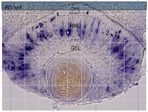
Cross-section of the eye after in situ hybridization using a ptf1a antisense probe (see Lin et al., Dev. Biol. 274, 491-503 (2004) for Methods). Arrows, location of outer plexiform layer; ONL, outer nuclear layer; INL, inner nuclear layer; GCL, ganglion cell layer. Asterisk: labeled cell in amacrine cell layer. (1,2) Labeled cells in outer half of INL. EXPRESSION / LABELING:
|