- Title
-
Isolation and expression of zebrafish zinc-finger transcription factor gene tsh1
- Authors
- Wang, H., Lee, E.M., Sperber, S.M., Lin, S., Ekker, M., and Long, Q.
- Source
- Full text @ Gene Expr. Patterns
|
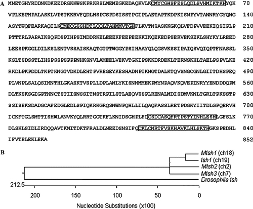
The zebrafish tsh1 gene encodes a zinc-finger transcription factor related to the Drosophila tsh. (A) Amino acid sequence of Tsh1. The zinc-finger motifs are boxed. (B) Phylogenetic tree of Drosophila tsh, zebrafish tsh1 and three mouse Tsh-like genes. The chromosomal location of each gene is indicated in parentheses. The phylogenetic tree was generated by use of Megalign of Lasergene 6 (DNAStar). The GenBank accession number for tsh, Mtsh1, Mtsh2 and Mtsh3 are: AAA28983, XM_888252, NM_080455, NM_172298, respectively. The scale at the bottom indicates total nucleotide substitutions between compared sequences. |
|
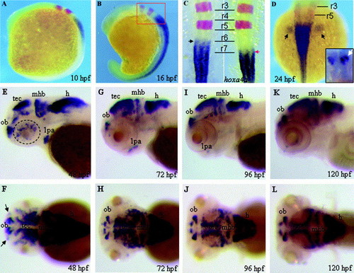
Spatial and temporal expression patterns of tsh1 revealed by RNA in situ hybridization. (A–D) Double in situ hybridization with tsh1 (blue) and krox-20 (red) cRNA probes. (E–L) Single in situ hybridization with tsh1 cRNA probe. The developmental stages of the hybridized embryos are indicated as hours post-fertilization (hpf). (A, B, E, G, I and K) are lateral views and (C, D, F, H, J and L) are dorsal views of the hybridized embryos. The inset in (D) is an anterior view (dorsal to the top) of the forebrain of the hybridized embryo. (A) At the 2-somite stage (10 hpf), tsh1 expression is initiated in the hindbrain and anterior spinal cord. (B) At the 14-somite stage (16 hpf), tsh1 is expressed throughout the spinal cord. (C) Dorsal view of the hindbrain region (red box in B). The expression domain of tsh1 has a clear anterior boundary at the rostral margin of rhombomere 7 (r7). (D) At the prim-5 stage (24 hpf), tsh1 is expressed in the spinal cord and pectoral fin buds (black arrows) and in the dorsal forebrain (white arrow in the inset). (E–F) At the long-pec stage (48 hpf), tsh1 is expressed in the olfactory bulb (ob), tectum opticum (tec), mid–hindbrain boundary (mhb), hindbrain (h), first pharyngeal arch (1 pa) and in the eye (dash line-circled), with very weak expression in the olfactory placodes (arrows in F). (G–H) At the protruding mouth stage (72 hpf), lower levels of tsh1 transcripts are observed in the olfactory bulb, pharyngeal arch and midbrain–hindbrain boundary. (I–L) At the early larvae stages (96–120 hpf), reduced expression of tsh1 is seen in the tectum and first arch. By 120 hpf, no tsh1 expression is detectable in the pharyngeal arches, but is still observed in restricted areas of the telecephalon, tectum, mid–hindbrain boundary and hindbrain. EXPRESSION / LABELING:
|
|
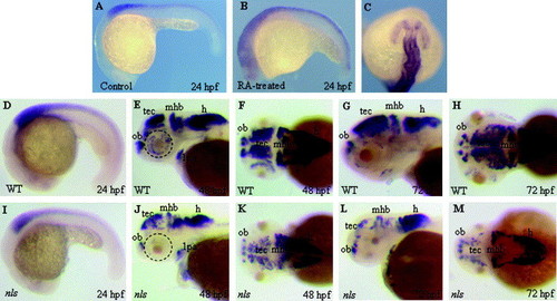
Expression of tsh1 is affected by retinoic acid signaling. (A–C) Expression of tsh1 expression in mock (A) and RA (B–C) treated embryos. (C) A dorsal view (anterior to the top) of the embryo in (B). (D, E, F, G and H) Lateral or dorsal view of tsh1 expression in wild-type and (I, J, K, L and M) neckless (nls) mutants. At 24 hpf, tsh1 expression in the spinal cord is markedly reduced in the nls mutant embryo (I) compared to wild-type siblings (D). Between 48 and 72 hpf, nls mutants (J, K, L and M) exhibit reduced tsh1 expression in the tectum, eyes, the midbrain–hindbrain boundary and the hindbrain. Abbreviations: mhb, mid–hindbrain; tec, tectum opticum; t, telencephalon; ob, olfactory bulb; h, hindbrain. EXPRESSION / LABELING:
|
Reprinted from Gene expression patterns : GEP, 7(3), Wang, H., Lee, E.M., Sperber, S.M., Lin, S., Ekker, M., and Long, Q., Isolation and expression of zebrafish zinc-finger transcription factor gene tsh1, 318-322, Copyright (2007) with permission from Elsevier. Full text @ Gene Expr. Patterns


