Lab
Chien Lab
|

|
Statement of Research Interest
The lab's overall interest is in how cell movements are controlled in the context of an intact, developing organism.
Axon guidance
Our largest effort focuses on how the growth cone guides the growing axon as it navigates through the brain, using the retinotectal system as a model. We use four different approaches, with the long-term aim of elucidating all of the genes and molecules that help to guide retinal axons to their targets, and understanding their functions in vivo.
1) Understanding the mutants. Several years ago we cloned the astray mutant and showed that it is defective in Robo2, a guidance receptor expressed in retinal ganglion cells (RGCs). We have characterized in detail the cell biological function of this receptor and its presumed ligands, the Slits. We have also cloned the boxer and dackel mutants (in collaboration with Henry Roehl), and found that they are defective in heparan sulfate proteoglycan (HSPG) synthesis. We are now working to define exactly how and when HSPGs act in optic tract sorting. Finally, we have cloned the nevermind mutant, defective in the cyfip2 gene, which encodes a protein in the WAVE complex that also binds to FMR1 family members.
2) Understanding the roles of candidate pathfinding molecules. Homologs of several molecules including Roundabout, neogenin, neuropilin, DCC, and unc5 are expressed in RGCs. We are developing techniques to transiently express wildtype and mutated forms of these molecules in RGCs, to test their role in retinal axon guidance.
3) Understanding how growth cones behave in vivo. We have developed techniques to make in vivo timelapse movies of navigating retinal growth cones, and are characterizing their dynamic behavior in both mutant and wildtype embryos. We have also developed several transgenic lines that express fluorescent protein markers in retinal axons.
4) Screening for new genes. We have carried out a large-scale noncomplementation screen for new robo2 alleles, and have an ongoing screen to look for mutants defective in early eye morphogenesis and patterning.
Cell motility in other contexts
Other projects in the lab study vascular guidance, dorsoventral eye patterning, and eye morphogenesis.
5) In the vascular system, we have been studying the role of the Netrin1a pathway in guiding secondary intersegmental sprouts to turn laterally and then anteroposteriorly at the horizontal myoseptum. These "parachordal" cells are of particular interest since they are the precursors of the zebrafish's lymphatic system.
6) We are studying the roles of the Bmp, Wnt, and Tbx pathways in controlling dorsoventral identity in the eye: initiation, maintenance, and refinement of dorsal identity, and eventually topographic axon guidance.
7) We have used 4D in toto imaging to visualize the development of the optic vesicle between 12-24 hpf, and have discovered several unappreciated cell movements, as well as defining fate maps of the eye's three main components: neural retina, retinal pigment epithelium, and lens.
Common resources
Finally, we are heavily involved in developing community resources:
8) To easily manipulate gene expression and generate transgenic lines, we developed the Tol2kit set of plasmids for multisite Gateway cloning of Tol2 expression constructs.
9) To visualize multichannel confocal datasets (both 3D and 4D), we have collaborated with Chuck Hansen's group in the University of Utah's Scientific Computing Institute to develop FluoRender, a freely-distributed volume rendering program, optimized to deliver useful visualizations quickly and with precision.
10) We are currently conducting a Gal4 enhancer trap screen, for which we are documenting expression patterns by widefield and confocal microscopy; lines will be distributed through ZIRC. We have discovered many beautiful patterns, including ones that label specific cells in the skin, lateral line, and nervous system. This work is funded by the NIMH under the rubric of the Zebrafish Tools initiative.
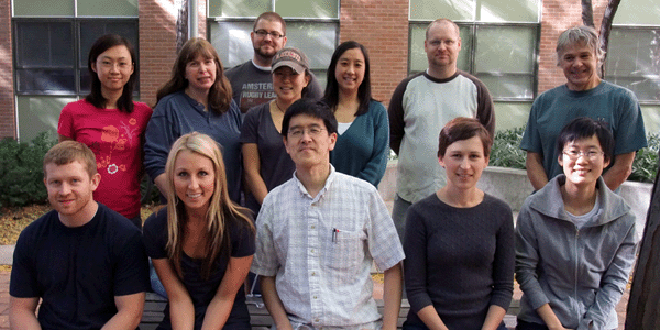
Lab Members
| Otsuna, Hideo Post-Doc | Bend, Renee Graduate Student | Gaynes, John Graduate Student |
| Hörndli, Coni Stacher Graduate Student | Law, Mei-Yee Graduate Student | Lim, Amy Graduate Student |
| Rasband, Kendall Graduate Student | Benko, Kevin Technical Staff | Fujimoto, Esther Technical Staff |
| Gaynes, Brooke Technical Staff | Quist, Tyler Technical Staff | Rosenthal, Jude Technical Staff |
| Scoresby, Aaron Technical Staff |
