
|
Fig. 1
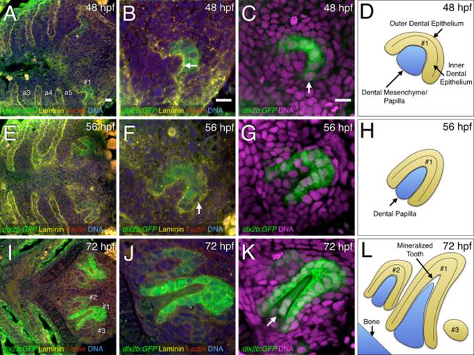
Fluorescence visualization of cellular details during tooth germ morphogenesis. A,E,I: Ventral views, anterior to the left, of the zebrafish pharyngeal region. B,C,F,G,J,K: Closeups of right-side tooth germs. A,B,E,F,I,J: Four-color stains of dlx2b:GFP reporter expression (green), laminin protein (yellow), f-actin (red), and DNA (blue). C,G,K: Two-color stains with dlx2b:GFP (green) and DNA (magenta). A,B: Laminin expression at 48 hpf and 56 hpf stages at the boundaries of the pharyngeal arches (a3–a5) as well as the interface between the dental epithelium and dental mesenchyme of tooth germ #1 (B, arrow). C: dlx2b:GFP reporter expression mostly in the inner dental epithelium (arrow). D: Schematic drawing of a early morphogenesis stage tooth germ #1 at 48 hpf. E,F: Laminin expression highlighting the outer dental epithelium in a late morphogenesis stage tooth germ #1 at 56 hpf (F, arrow). G,H: dlx2b:GFP expression in the inner dental epithelium and schematic drawing. I,J: F-actin staining along with dlx2b:GFP expression in cell differentiation stage tooth germ #1 at 72 hpf. Tooth germs #2 and #3 are also present. K: dlx2b:GFP expression in the inner dental epithelium and the dental papilla (arrow) at 72 hpf. Tooth germs #2 and #3 are mostly outside the focal plane in this specimen. L: Schematic drawing of typical orientations of the tooth germs at 72 hpf. Scale bars = 10 µm in A–C.