
Image
|
Figure Caption
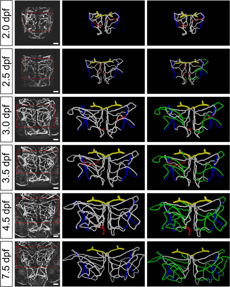
Fig. S2 Developmental expansion of midbrain vasculature during 2.0 to 7.5 dpf. Projected confocal images (left) and 3-D reconstruction (middle, right) of a larva′s midbrain vasculature imaged at 2.0, 2.5, 3.0, 3.5, 4.5, and 7.5 dpf. The dashed square delineates the midbrain position. The segments that were pruned at the next time point are marked in red (middle). The newly formed segments after 2.0 dpf through angiogenesis are marked in green (right). Yellow, BCA; white, midbrain vasculature; blue, CVP. Scale, 50 μm.
Figure Data
Acknowledgments
This image is the copyrighted work of the attributed author or publisher, and
ZFIN has permission only to display this image to its users.
Additional permissions should be obtained from the applicable author or publisher of the image.
Full text @ PLoS Biol.