
Image
|
Figure Caption
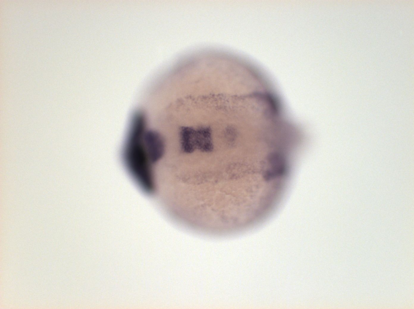
Fig. 3 forebrain, retina, ventral posterior diencephalon, ventral hindbrain stronger in rhombomeres 1, 2, 3, (not in 4), weak in 5, ventral cephalic mesenchyme, weak in YSL
Orientation
| Preparation | Image Form | View | Direction |
| whole-mount | still | dorsal | anterior to left |
Figure Data